

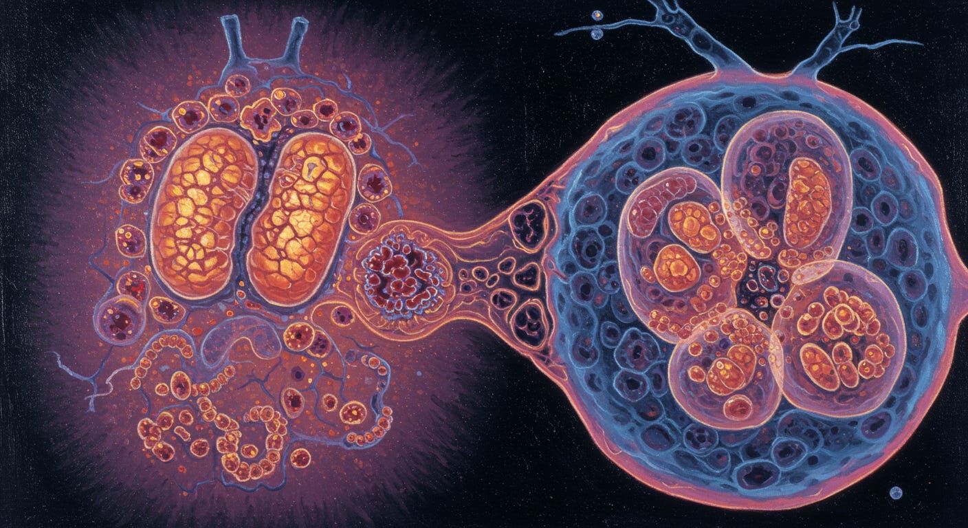
Cancer is not merely a disease but a multi-trillion-dollar industrial enterprise, a mechanism that transfers wealth from patients to a vast medical-industrial complex in their final days. The conventional "standard of care"—a triad of poisoning with chemotherapy, burning with radiation, and cutting with surgery—dominates oncology, yet it’s a limited, dehumanizing approach. Tripping over the Truth outlines the metabolic theory of cancer, which challenges the genetic narrative. This view, rooted in Otto Warburg’s 1924 observation of the Warburg effect—where cancer cells ferment glucose even in oxygen-rich environments—suggests that cancer stems from mitochondrial dysfunction, not DNA mutations. Evidence from nuclear transfer experiments, as noted in Seyfried’s Cancer as a Metabolic Disease, reinforces this: cancer persists in cells with normal nuclei if the cytoplasm is damaged. Skepticism of the industrial cancer narrative, critiqued in Chemotherapy, stems from its profit-driven focus on genetic therapies over addressing these metabolic roots.
Historically, the somatic mutation theory (SMT) has reigned since the 1970s, propelled by discoveries like oncogenes and ambitious efforts such as The Cancer Genome Atlas (TCGA). Yet, TCGA revealed a chaotic landscape of genetic heterogeneity—some cancers lack clear driver mutations, a puzzle dubbed the “dark matter” of cancer, as explored in Cowan’s Cancer and the New Biology of Water. The metabolic theory offers a counterpoint: cancer’s universal reliance on fermentation, visualized via PET scans, points to energy metabolism as the primary defect. Nuclear transfer experiments further undermine SMT—transplanting a cancer nucleus into healthy cytoplasm rarely produces tumors, while the reverse almost always does, per The Top 10 Cancer Cures No One Is Talking About. This shift in understanding, from genes to mitochondria, resonates with the broader rejection of allopathic paradigms and rigidity. It’s a lens that aligns with my belief that we must educate ourselves beyond the narratives upheld by a system that, as A New Standard of Care Documentary reveals, prioritizes profit over paradigm shifts.
The promise of metabolic therapies are approaches like the ketogenic diet and 3-bromopyruvate (3BP) target cancer’s metabolic Achilles’ heel. The ketogenic diet, detailed in Radical Remission and The Gerson Therapy, starves cancer of glucose, leveraging its inability to metabolize ketones efficiently. Meanwhile, 3BP, a glycolysis inhibitor, has shown striking preclinical success, yet remains stifled by regulatory and industry barriers, as noted in Cancer Care. These alternatives face resistance from a cancer establishment that favors patentable drugs over affordable, non-patentable solutions. Clinics like ChemoThermia Oncology Center demonstrate that metabolically supported treatments can outperform conventional methods, yet adoption lags. We must take responsibility for our education, rejecting the industrial cancer narrative to embrace a future where healing, not business, drives care.
With thanks to Travis Christofferson.
Deep Dive Conversation Library (Bonus for Paid Subscribers Only)
This deep dive is based on the book:
Discussion No.80:
23 insights and reflections from “Tripping over the Truth”
Thank you for your support.
Analogy
The Analogy of the Car Factory
Imagine cancer as a problem in a car factory. For decades, experts believed the factory was malfunctioning because of errors in the engineering blueprints (the DNA). They devoted enormous resources to studying these blueprints, cataloging every possible mistake, and developing specialized tools to fix each specific blueprint error. Yet despite identifying thousands of blueprint flaws and creating hundreds of specialized repair tools, most of the factory's problems persisted.
What if the real issue wasn't primarily in the blueprints, but in the factory's power plant? Imagine that damaged power generators had been forcing the factory to run on an emergency backup system – an old, inefficient diesel generator instead of the modern electrical grid. This emergency power system was meant only for temporary crises, but when the main power plant became permanently damaged, the backup never switched off.
Running continuously on this emergency power creates numerous problems: it's inefficient, requiring massive amounts of fuel (glucose); it produces excessive pollution (lactic acid); and most importantly, the inconsistent power supply causes the blueprint-reading machines to make errors, introducing flaws into the manufacturing process. The longer the factory runs on emergency power, the more blueprint errors accumulate.
The metabolic theory of cancer suggests we've been focusing on fixing thousands of different blueprint errors, when we could instead repair the power plant or cut off fuel to the emergency generator. This approach might work regardless of which specific blueprint errors have accumulated in a particular factory.
This perspective explains why some approaches that don't directly target blueprint errors – like modified diets that restrict the fuel (glucose) needed by the emergency generator, or compounds that disable the emergency power system – have shown promise across many different types of cancer, despite their varied genetic profiles. It suggests we might have been tripping over the truth all along: the power problem comes first, and the blueprint errors follow.
12-point summary
1. Competing Theories of Cancer Origin: The book presents two competing theories of cancer causation. The Somatic Mutation Theory (SMT) proposes that cancer is caused by sequential mutations to DNA, with genetic damage as the primary driver. The Metabolic Theory, first proposed by Otto Warburg in 1924, suggests that cancer originates from damaged mitochondria and impaired cellular respiration, forcing cells to rely on fermentation metabolism even in the presence of oxygen (the Warburg effect). This metabolic shift then triggers genetic instability and mutations as secondary effects rather than primary causes.
2. Historical Development of Cancer Research: Cancer research has evolved through distinct phases, beginning with Percivall Pott's 1775 discovery linking environmental carcinogens to cancer. David von Hansemann later observed chaotic chromosomes in cancer cells, and Peyton Rous discovered viral causes of cancer. Watson and Crick's DNA structure discovery in 1953 shifted focus toward genetic explanations. The pivotal moment came in 1976 when Harold Varmus and Michael Bishop discovered oncogenes, cementing the genetic theory as dominant for decades until genomic sequencing projects revealed unexpected complexity that challenged this paradigm.
3. The Warburg Effect and Cancer Metabolism: All cancer cells exhibit a peculiar metabolic pattern called the Warburg effect—they generate energy through glucose fermentation even in the presence of oxygen, a highly inefficient process requiring 18 times more glucose than normal aerobic respiration. This universal feature appears across all cancer types regardless of their genetic mutations. PET scans, which detect areas of high glucose consumption, visualize this effect and have become the gold standard for cancer detection, ironically making practical use of Warburg's theory even when it was scientifically marginalized.
4. Mitochondrial Damage and Retrograde Response: The metabolic theory centers on mitochondria—cellular "power plants" that generate energy through respiration. Cancer cells typically have fewer mitochondria, and those remaining show structural abnormalities. When mitochondria become damaged, they send distress signals to the nucleus through the "retrograde response," activating genes that enable fermentation metabolism and cellular survival. This response also activates oncogenes controlling cell proliferation while reducing DNA repair mechanisms, explaining the genetic instability and mutations observed in cancer cells.
5. The Cancer Genome Atlas Revelations: The Cancer Genome Atlas project, launched in 2006 to catalog all cancer-causing mutations, produced unexpected results that challenged the genetic theory. Rather than revealing consistent mutational patterns for each cancer type, it exposed tremendous heterogeneity—mutations varied dramatically between patients with the same cancer (intertumoral heterogeneity) and even between cells within the same tumor (intratumoral heterogeneity). Some aggressive cancers showed few or no driver mutations, leading researcher Bert Vogelstein to propose the existence of cancer "dark matter"—unknown factors driving cancer beyond detectable mutations.
6. Nuclear Transfer Experiments: Perhaps the most compelling evidence supporting the metabolic theory came from nuclear transfer experiments conducted independently by Warren Schaeffer and Jerry Shay in the 1980s. When they transplanted the nucleus of a cancer cell (containing all its mutated DNA) into a normal cell with its nucleus removed, the resulting cells rarely formed tumors. Conversely, when placing normal nuclei into cancer cell cytoplasm, 97% of cases became cancerous. These findings suggested that something in the cytoplasm—where mitochondria reside—was more determinative of cancer than nuclear DNA, directly challenging the genetic theory.
7. Evolution of Chemotherapy: The first chemotherapy agent originated from a World War II accident when mustard gas exposure was observed to deplete white blood cells. This led to nitrogen mustard's development for treating lymphoma, though initial remissions proved brief. Pioneers like Emil Frei, Emil Freireich, Vincent DeVita, and Donald Pinkel developed increasingly effective protocols combining multiple drugs (VAMP, MOPP, Total Therapy) to prevent resistance, establishing principles still used today. Although effective for certain cancers like leukemia, Hodgkin's disease, and testicular cancer, these toxic approaches failed to significantly impact most common cancers, with overall cancer death rates remaining largely unchanged since the 1950s.
8. Targeted Therapy Limitations: The genetic theory of cancer led to the development of targeted therapies designed to attack specific mutations. Trastuzumab (Herceptin) and imatinib (Gleevec) became celebrated examples, but their success proved limited. Herceptin extended life by only about four months in metastatic breast cancer and worked for only 20% of breast cancer patients. Gleevec was more successful for chronic myeloid leukemia but proved an outlier due to that cancer's unique genetic homogeneity. The vast genetic heterogeneity revealed by genome sequencing explained why most targeted therapies failed—there was simply no consistent mutational target across patients with the same cancer type.
9. 3-Bromopyruvate Discovery and Potential: In 2000, Young Hee Ko in Peter Pedersen's laboratory discovered 3-bromopyruvate (3BP), a molecule that targets cancer's metabolic vulnerability. 3BP enters cancer cells through their overexpressed monocarboxylate transporters and inhibits hexokinase II, cutting off cancer cells' energy supply. In laboratory and animal studies, 3BP showed remarkable efficacy, completely eradicating advanced liver cancers in rats without harming normal tissues. The first human case, Dutch teenager Yvar Verhoeven with terminal liver cancer, showed complete tumor regression after 3BP treatment, though he later died from pneumonia complications. Despite this promise, 3BP's development has been hampered by controversies, patent disputes, and regulatory challenges.
10. Ketogenic Diet as Cancer Therapy: The ketogenic diet (high fat, adequate protein, very low carbohydrate) forces the body to produce ketone bodies instead of relying on glucose. Since cancer cells typically cannot efficiently use ketones due to their damaged mitochondria, this creates a selective metabolic pressure. Linda Nebeling's pioneering 1995 study showed the diet reduced glucose uptake in brain tumors by 22%, with patients surviving years beyond their prognoses. Later case studies, like Marianne Zuccoli's glioblastoma remission while on the restricted ketogenic diet, provided further evidence. The diet appears to work both as a standalone therapy and as an enhancer of conventional treatments, improving efficacy while reducing side effects.
11. Press-Pulse Approach to Cancer Treatment: Thomas Seyfried and Dominic D'Agostino developed the "press-pulse" strategy for cancer treatment, inspired by ecological concepts of mass extinction. The "press" involves sustained metabolic stress through caloric restriction and ketogenic diet, while "pulses" consist of acute interventions like metabolic drugs (3BP, 2-deoxyglucose) or hyperbaric oxygen therapy. When combined, these approaches show remarkable synergy—mice with metastatic cancer showed 77.9% increased survival with ketogenic diet plus hyperbaric oxygen versus either treatment alone. ChemoThermia Oncology Center in Istanbul has applied a similar approach in humans, reporting dramatic improvements in survival for pancreatic and lung cancers compared to conventional treatment alone.
12. Regulatory and Research Challenges: Despite promising results from metabolic approaches, several barriers impede their development. Because metabolic treatments are often inexpensive (especially dietary approaches) or use off-patent compounds, pharmaceutical companies have little financial incentive to fund costly clinical trials. The regulatory system is structured to test single agents sequentially rather than combinations, though cancer likely requires multi-targeted approaches. Some researchers advocate for learning from the AIDS activist model, where doctors and patients combined existing drugs out of necessity, ultimately developing life-saving cocktail therapies without going through traditional regulatory channels. Care Oncology Clinic, Notable Labs, and ChemoThermia represent pioneering efforts to test such approaches outside conventional research paradigms.
45 Questions and Answers
Question 1: What is the Warburg effect and how does it relate to cancer metabolism?
The Warburg effect describes a peculiar metabolic pattern observed in cancer cells where they generate energy through glucose fermentation even in the presence of oxygen. Unlike normal cells that primarily rely on the efficient process of aerobic respiration using oxygen to produce energy, cancer cells shift to the highly inefficient pathway of fermentation. This phenomenon was first documented by Otto Warburg in 1924, who noticed that cancer cells produced abnormal amounts of lactic acid, indicating they were fermenting glucose instead of fully metabolizing it through oxidative respiration. The process is remarkably inefficient—generating energy through fermentation requires about eighteen times more glucose to extract the same amount of energy as aerobic respiration.
This metabolic abnormality has profound implications for understanding cancer. Warburg eventually concluded this was not merely a side effect but the actual origin of cancer, stating "the prime cause of cancer is the replacement of the respiration of oxygen in normal body cells by a fermentation of sugar." Modern research has confirmed this metabolic pattern exists across virtually all cancer types, making it a universal feature detectable through PET scanning. The Warburg effect suggests cancer cells have damaged mitochondria (the cellular power plants), forcing them to compensate through increased fermentation. This creates a distinctive metabolic vulnerability that researchers like Peter Pedersen, Young Ko, and Thomas Seyfried have sought to exploit therapeutically.
Question 2: How did Otto Warburg's theory about cancer differ from the Somatic Mutation Theory?
Warburg's theory proposed that cancer originates from damage to cellular respiration, specifically the cell's ability to generate energy through oxidative processes in the mitochondria. He contended that this respiratory damage occurs first, causing cells to shift to fermentation metabolism even in the presence of oxygen, which he considered the defining characteristic of cancer cells. Warburg believed this metabolic shift was the "prime cause" into which all secondary causes (carcinogens, viruses, radiation) converged. According to him, the metabolic alteration was not just a symptom but the fundamental event that initiated the transformation from normal to malignant cells.
In stark contrast, the Somatic Mutation Theory (SMT) holds that cancer is caused by sequential mutations to DNA, particularly to oncogenes and tumor suppressor genes. The SMT proposes that these genetic changes rewire critical cellular circuitry, progressively transforming a cell toward malignancy. While Warburg considered genomic instability and mutations to be secondary effects of damaged metabolism, the SMT positions them as the primary drivers of cancer. This fundamental difference in causal direction creates two competing paradigms: the metabolic theory sees mutations as side effects of mitochondrial damage, while the genetic theory views metabolic changes as downstream consequences of genetic alterations. The triumph of the SMT throughout most of the 20th century effectively sidelined Warburg's metabolic theory until its recent resurgence.
Question 3: What was Percivall Pott's contribution to cancer research?
Percivall Pott made a groundbreaking contribution to cancer research in 1775 when he established the first documented link between an environmental agent and cancer. As a surgeon in London, Pott noticed an unusual prevalence of scrotal cancer among chimney sweeps, particularly young boys who had worked as chimney sweeps from an early age. Through careful observation, he identified that the "soot warts" commonly seen on these boys' scrotums were actually a rare form of cancerous growth, and he theorized that prolonged exposure to chimney soot was the causative factor.
Pott's observation was revolutionary because it connected an external agent to cancer development, introducing the concept that would later be known as carcinogenesis. His work established that cancer could be caused by environmental factors, laying the groundwork for the field of occupational carcinogenesis. This discovery shifted medical thinking about cancer from viewing it as an internal imbalance to recognizing external influences in its development. Pott's work became the foundation for the growing list of cancer-causing agents (carcinogens) identified over subsequent centuries and formed a key building block of what would later become the Somatic Mutation Theory, as it suggested external agents could alter something critical within cells to cause uncontrolled growth.
Question 4: How did the discovery of the Rous sarcoma virus change cancer research?
The discovery of the Rous sarcoma virus by Peyton Rous in 1910 dramatically altered the cancer research landscape by demonstrating that cancer could have a viral origin. Rous showed that a solid tumor (a spindle cell sarcoma) in chickens could be transmitted to other chickens not just by transplanting tumor tissue, but also by injecting filtered tumor material that contained no actual cancer cells—only what we now know was the virus. This definitively proved that some cancers could be caused by infectious agents, a concept that was revolutionary at the time.
This discovery complicated the emerging narrative about cancer causation. While Pott had established environmental carcinogens as a cause, and Hansemann had observed chromosomal abnormalities in cancer cells, Rous's viral theory introduced a third, seemingly unrelated mechanism. The Rous sarcoma virus discovery prevented the formation of a single, comprehensive theory on cancer's origin for most of the 20th century. It wasn't until Varmus and Bishop's Nobel Prize-winning work in 1976 that Rous's viral theory was reconciled with the Somatic Mutation Theory, when they showed that the virus contained a slightly altered version of a normal cellular gene. This integration of theories helped cement the SMT as the dominant paradigm, though ironically, later evidence would show that the Rous virus also directly damaged mitochondria, potentially supporting Warburg's metabolic theory as well.
Question 5: What is the Somatic Mutation Theory (SMT) of cancer and how did it develop?
The Somatic Mutation Theory proposes that cancer originates from accumulated mutations in the DNA of somatic (body) cells, which progressively transform normal cells into malignant ones. These mutations typically affect proto-oncogenes (which can become cancer-promoting oncogenes) and tumor suppressor genes, resulting in uncontrolled cell proliferation, invasion, and metastasis. The theory suggests that cancer develops through a series of genetic alterations that accumulate over time, each conferring growth advantages to the affected cells, ultimately leading to full malignancy.
The SMT developed gradually through multiple discoveries. It began with Pott's identification of environmental carcinogens in the 1700s and Hansemann's observations of chaotic chromosomes in cancer cells in the 1890s. The theory gained momentum when Watson and Crick discovered DNA's structure in 1953, providing a molecular basis for genetic inheritance and mutation. The definitive breakthrough came in 1976 when Varmus and Bishop discovered that the cancer-causing gene in the Rous sarcoma virus was actually a mutated version of a normal cellular gene. This tied together environmental carcinogens, viral carcinogenesis, and chromosomal abnormalities into a unified theory: all could cause cancer by mutating critical genes. The SMT became the dominant paradigm, guiding cancer research for decades and inspiring massive projects like The Cancer Genome Atlas, which sought to catalog all cancer-causing mutations.
Question 6: What role do mitochondria play in the metabolic theory of cancer?
In the metabolic theory of cancer, mitochondria take center stage as the primary site of cancer initiation. The theory proposes that damage to these cellular "power plants" forces cells to shift from efficient aerobic energy production to inefficient fermentation metabolism (the Warburg effect). According to researchers like Pedersen and Seyfried, cancer cells typically have fewer mitochondria, and those that remain show structural abnormalities—they appear smaller, misshapen, and lacking important internal membranes. This damage compromises the cell's ability to generate energy through oxidative phosphorylation, creating a metabolic crisis that triggers emergency compensatory mechanisms.
When mitochondria become damaged beyond a critical threshold, they send distress signals called the "retrograde response" to the nucleus, activating genes that enable fermentation metabolism and cellular survival under impaired respiration. This response also activates oncogenes like MYC, RAS, and others that control cell proliferation and survival. The theory proposes that this mitochondrial-to-nuclear signaling changes gene expression patterns throughout the cell, ultimately manifesting as cancer's hallmark features. Importantly, the metabolic theory turns the SMT's causal direction upside-down: rather than mutations causing cancer, the theory suggests that mitochondrial damage precedes and actually causes the genomic instability and mutations observed in cancer cells. This perspective explains why cancer cells universally exhibit the Warburg effect regardless of their tissue of origin or specific mutations.
Question 7: What were the key discoveries of Harold Varmus and Michael Bishop regarding oncogenes?
Harold Varmus and Michael Bishop made the pivotal discovery that cancer-causing genes (oncogenes) in viruses were actually altered versions of normal genes present in all animals. In 1976, they isolated the src gene from the Rous sarcoma virus, which was known to cause cancer in chickens. Using molecular "fishing" techniques, they discovered that similar genes existed in normal cells of birds, mammals, fish, and even humans. This meant that the cancer-causing viral genes weren't foreign invaders but modified versions of our own genetic material.
Their breakthrough revealed that the viral src gene differed from normal cellular SRC by only a few nucleotides, but these small changes were enough to create a permanently activated protein that continuously signaled cells to divide. This discovery unified previously competing theories of cancer causation by showing that carcinogens, radiation, and viruses could all cause cancer through the same mechanism: by altering proto-oncogenes into oncogenes. Varmus and Bishop's work established that cancer was fundamentally a genetic disease, as they demonstrated how a single altered gene could transform cellular behavior. Their discovery was so influential it won them the Nobel Prize and cemented the Somatic Mutation Theory as the dominant paradigm for decades, guiding research efforts like targeted drug development and The Cancer Genome Atlas project.
Question 8: How did the discovery of DNA influence cancer research?
The discovery of DNA's double helix structure by Watson and Crick in 1953 fundamentally transformed cancer research by providing the molecular framework to understand how genetic information is stored and transmitted. This breakthrough shifted scientific thinking about disease causation toward genetic mechanisms and away from other theories, including Warburg's metabolic theory. Once scientists understood that DNA contained the instructions for all cellular processes, it became logical to search for the origin of cancer within this genetic blueprint. The discovery initiated what the New York Times called "the golden age of molecular biology," during which the genetic code and protein synthesis were deciphered.
With DNA established as the central molecule of life, cancer researchers increasingly focused on identifying mutations that could convert normal cells to cancerous ones. By the 1960s, the idea that DNA alterations were central to cancer was widely accepted, as articulated by Frank Horsfall in 1963: "Because the cancerous change in cells appears to be a permanent alteration, handed on to daughter cells through innumerable divisions, it seems probable that it reflects an abnormality in the transfer of information from cell to daughter cells." This genetic perspective provided the conceptual foundation for later discoveries by Varmus and Bishop, who identified specific oncogenes, and ultimately led to the development of targeted cancer therapies designed to counteract the effects of specific genetic mutations.
Question 9: What is 3-bromopyruvate (3BP) and how was it discovered?
3-bromopyruvate (3BP) is a small molecule that functions as a potent metabolic inhibitor targeting cancer cells' energy production mechanisms. Structurally, it resembles pyruvate (a key metabolic intermediate) but contains a bromine atom that makes it highly reactive. 3BP exploits cancer cells' altered metabolism by entering through their overexpressed monocarboxylate transporters (MCTs) and then inhibiting hexokinase II, the enzyme that initiates glucose fermentation and is critical for cancer cells' survival. It effectively cuts off cancer cells' energy supply by blocking both fermentation and oxidative pathways simultaneously, causing rapid energy depletion and cell death.
3BP was discovered by Young Hee Ko in Peter Pedersen's laboratory at Johns Hopkins University around 2000. Ko had been working on cystic fibrosis research but shifted to cancer metabolism when she joined Pedersen's team. Having observed that hexokinase II was overexpressed in cancer cells and bound to mitochondria, Ko hypothesized that a molecule resembling pyruvate but with a reactive element might disrupt this critical energy-generating system. Drawing on her background in biochemistry, she selected 3BP from chemical supply catalogs and tested it against cancer cells. The results were stunning—3BP killed cancer cells more effectively than conventional chemotherapy drugs in petri dish experiments. Despite initial skepticism about its reactivity, subsequent tests in animals showed that 3BP could eradicate advanced liver cancers without apparent toxicity to normal tissues, making it a potentially revolutionary cancer treatment.
Question 10: What was Yvar Verhoeven's experience with 3BP treatment?
Yvar Verhoeven, a teenage boy from the Netherlands, became the first documented human case successfully treated with 3BP after being diagnosed with advanced hepatocellular carcinoma in 2008. Initially presenting with persistent burping, Yvar was eventually found to have massive tumors consuming 95% of his liver with metastasis to his heart. After failing to respond to conventional treatments including sorafenib, his father Harrie Verhoeven discovered information about 3BP online and contacted Young Ko directly. With no other options remaining, Ko helped coordinate treatment with German physician Thomas Vogl at the University of Frankfurt.
Yvar received 3BP treatments via a catheter procedure called transcatheter arterial chemoembolization (TACE), which delivered the compound directly to his liver tumors. The results were remarkable—within hours of his first treatment, Yvar reported feeling hungry for the first time in months. After multiple treatments over several months, imaging showed complete regression of his tumors, with evidence of liver regeneration. Yvar recovered enough to travel, speak at medical conferences, and resume normal teenage activities. Though he ultimately succumbed to pneumonia when his weakened body couldn't process the necessary antibiotics, CT scans confirmed he was cancer-free at the time of his death. His case provided powerful evidence for 3BP's efficacy, though the compound's development would later be complicated by controversy and legal disputes.
Question 11: How did chemotherapy originate and develop as a cancer treatment?
Chemotherapy originated in a tragic accident during World War II. On December 3, 1943, German planes bombed the port of Bari in Italy, destroying several Allied ships including one carrying mustard gas. The gas spilled into the water, and victims experienced not only burns and blindness but also showed striking depletion of white blood cells in their lymph nodes and bone marrow. Lieutenant Colonel Stewart Alexander collected tissue samples from the victims, and these samples later inspired Yale pharmacologists Louis Goodman and Alfred Gilman to investigate whether the war gas might be used to treat lymphomas, which were characterized by excessive white blood cell production.
Their initial studies in mice showed promising results, leading to the first human trial of nitrogen mustard in a patient with non-Hodgkin's lymphoma. The patient's tumors dramatically regressed, prompting further treatments in other patients. Though these early remissions proved brief and incomplete, this wartime accident had launched the chemotherapy era. Over subsequent decades, researchers like Sidney Farber, Emil Frei, Emil Freireich, and Vincent DeVita developed increasingly effective drug combinations. Their work established foundational principles that still guide cancer treatment: combining multiple drugs with different mechanisms of action, maintaining dose intensity, and treating for prolonged periods to ensure eradication of every cancer cell. Though initially rudimentary and highly toxic, chemotherapy development marked the beginning of systemic approaches to cancer treatment beyond surgery and radiation.
Question 12: What were the VAMP and MOPP chemotherapy protocols, and why were they significant?
VAMP and MOPP were groundbreaking combination chemotherapy protocols developed in the 1960s that revolutionized cancer treatment. VAMP, developed by Emil Frei and Emil Freireich at the National Cancer Institute for treating childhood leukemia, combined four drugs: vincristine, amethopterin (methotrexate), mercaptopurine, and prednisone. This protocol emerged from the realization that cancer could develop resistance to single drugs, but attacking it from multiple angles simultaneously could prevent this resistance. Despite initial opposition from other researchers who considered it too toxic, VAMP produced dramatic remissions in children with leukemia, though many later relapsed when cancer cells found refuge in the central nervous system.
MOPP, developed by Vincent DeVita for Hodgkin's lymphoma, consisted of nitrogen mustard, Oncovin (vincristine), procarbazine, and prednisone. When implemented in 1964 for patients with advanced Hodgkin's disease, MOPP achieved unprecedented results—60% of patients were cured. Similarly, Donald Pinkel later developed "Total Therapy" for childhood leukemia, which included multiple drug combinations and preventative treatment of the central nervous system, achieving 80% cure rates. These protocols were significant because they established that cancers could be cured with drugs alone—a paradigm shift from the prevailing perception that cancer was inevitably fatal once it had spread beyond surgical or radiation boundaries. VAMP and MOPP validated the combination approach that remains fundamental to cancer treatment today, even though they caused severe side effects including nausea, immune suppression, hair loss, sterility, and later increased risk of secondary cancers.
Question 13: What is the ketogenic diet's proposed mechanism of action against cancer?
The ketogenic diet's proposed mechanism against cancer centers on exploiting the metabolic vulnerabilities of cancer cells. Since cancer cells depend heavily on glucose fermentation for energy (the Warburg effect) and typically have damaged mitochondria rendering them unable to efficiently use ketone bodies, the diet creates a hostile metabolic environment specifically for malignant cells. By severely restricting carbohydrates and moderating protein intake while increasing fat consumption, the diet drives down blood glucose levels and forces the body to produce ketone bodies as an alternative energy source. Normal cells readily adapt to using these ketone bodies through their healthy mitochondria, but cancer cells struggle to make this metabolic shift.
Beyond simply restricting cancer's preferred fuel, the ketogenic diet activates multiple anti-cancer mechanisms. It reduces insulin and insulin-like growth factor signaling that many tumors depend on for growth stimulation. The diet appears to be anti-angiogenic, inhibiting the formation of new blood vessels that tumors need for expansion. It demonstrates pro-apoptotic effects, encouraging programmed cell death in cancer cells while reducing inflammation that can drive tumor progression. Additionally, ketone bodies themselves may have direct anticancer properties and can enhance normal cells' ability to withstand oxidative stress through increased glutathione production, while paradoxically making cancer cells more vulnerable to oxidative therapies like radiation and chemotherapy. Thomas Seyfried has described the diet as the "press" in a "press-pulse" strategy that weakens cancer cells and makes them more susceptible to targeted "pulse" treatments.
Question 14: How did PET scans develop from cancer metabolism research?
PET (Positron Emission Tomography) scans developed directly from Peter Pedersen's groundbreaking discoveries about cancer metabolism in the 1970s. Pedersen and his student Ernesto Bustamante had identified that cancer cells overexpress a specific enzyme called hexokinase II, which traps glucose inside cancer cells by tagging it with a phosphate molecule. This distinctive metabolic feature provided the basis for what would become the most important cancer imaging technology in history. The nascent PET scanning technology of the 1970s was floundering because researchers lacked a compound that would concentrate specifically in diseased tissue to create contrast against normal tissue.
The hexokinase II discovery provided exactly what PET scanning needed—a biological mechanism that would trap labeled glucose preferentially in cancer cells. Researchers developed fluorodeoxyglucose (FDG), a glucose analog with a radioactive fluorine atom that hexokinase II would process similarly to regular glucose. When injected into a patient, cancer cells with their overexpressed hexokinase II would rapidly accumulate FDG, which would then be detected by the scanner. Pedersen recalled giving a seminar about hexokinase II at the NIH where Giovanni Di Chiro, who was developing PET technology, was in attendance. Shortly after, FDG-PET scanning emerged as the gold standard for detecting actively metabolizing tumors. This application of metabolic theory ironically became one of the most widely used diagnostic tools even during an era when the genetic theory of cancer dominated research, with oncologists worldwide visualizing the Warburg effect daily through PET scans.
Question 15: What is intertumoral and intratumoral heterogeneity, and why are they significant?
Intertumoral heterogeneity refers to the vast differences in mutations found between tumors of the same cancer type in different patients. The Cancer Genome Atlas (TCGA) revealed that two people with the same diagnosis (e.g., breast cancer) might have completely different sets of mutations driving their disease, with few commonalities. This heterogeneity means that the mutations causing a specific type of cancer vary dramatically from person to person, making it incredibly difficult to identify universal driver mutations or develop broadly effective targeted therapies. The randomness and unpredictability of these mutations challenged researchers' expectations of finding consistent mutational patterns for each cancer type.
Intratumoral heterogeneity describes the variation in mutations found within a single tumor—from one region to another or even cell to cell. Advanced sequencing technology revealed that tumors are not homogeneous masses of identical cells but rather mosaics containing multiple subpopulations with different mutations. This diversity makes treatment extremely challenging, as drugs targeting one mutation might eliminate certain cancer cells while leaving others unaffected. Charles Swanton, who pioneered research in this area, described the implications as "mind-blowing" and likened treating such tumors to "playing whack-a-mole." Both forms of heterogeneity severely undermine the prevailing somatic mutation theory and pose tremendous obstacles to targeted therapy approaches. They suggest that cancer might not be driven solely by specific mutations but by some other process—potentially lending support to the metabolic theory of cancer, which proposes a more universal mechanism that could explain why cancer cells might accumulate diverse mutations as a side effect rather than a cause of malignancy.
Question 16: What were the outcomes of The Cancer Genome Atlas (TCGA) project?
The Cancer Genome Atlas, launched in 2006 as the culmination of decades of genetic cancer research, produced results that shocked the scientific community. Rather than revealing clear patterns of driver mutations that could explain different cancer types, TCGA uncovered an unexpected degree of randomness and complexity. For each cancer type, researchers found tremendous variation in mutations between patients (intertumoral heterogeneity), with no single mutation or combination consistently required for disease initiation. Beyond a few commonly mutated genes like p53, the mutational landscape appeared largely chaotic. Some cancers displayed only one or two mutations, while others showed six or more, contradicting the theory that cancer requires multiple sequential mutations to develop.
More troubling still, some cancer samples showed no identifiable driver mutations at all, despite being aggressive malignancies. These findings, described as "sobering" by scientists involved, significantly undermined the prevailing somatic mutation theory. Bert Vogelstein, a pioneer of the genetic approach, acknowledged this challenge by proposing the existence of cancer "dark matter"—unknown factors driving cancer beyond detectable mutations. The TCGA results had profound therapeutic implications, suggesting that the "targeted therapy" approach might be fundamentally limited because there was no consistent mutational target across patients. This led to a period of rethinking in cancer biology, with some researchers abandoning the genetic approach, others modifying the theory to account for "systems" dysfunction rather than specific mutations, and a small minority like Thomas Seyfried seeing the data as vindication of the metabolic theory of cancer.
Question 17: What is the "dark matter" in cancer that Bert Vogelstein referred to?
The term "dark matter" was borrowed by Bert Vogelstein from astrophysics to describe the mysterious drivers of cancer that remained undetected by genome sequencing. Just as dark matter in physics accounts for missing mass in the universe that can't be directly observed, cancer dark matter represented the unknown factors causing cancer that weren't visible in the genetic data. Vogelstein invoked this concept to explain a troubling discrepancy revealed by The Cancer Genome Atlas: many cancers showed far fewer driver mutations than expected. Some pediatric tumors had zero to two driver mutations, while many adult tumors had only three to six – numbers insufficient to explain the multiple hallmarks of cancer that were presumed to require sequential genetic alterations.
Vogelstein directly asked, "Where are these missing mutations?" The most likely candidate for this dark matter, he suggested, was epigenetic changes – alterations that affect gene expression without changing the DNA sequence itself. These might include DNA methylation patterns, histone modifications, and other regulatory mechanisms that control which genes are active or silent. From the perspective of metabolic theory proponents, this "dark matter" could be explained by mitochondrial dysfunction and the subsequent retrograde response, which would alter cellular function through epigenetic changes rather than direct DNA mutations. The concept of dark matter represented an acknowledgment that the somatic mutation theory, despite decades of dominance, couldn't fully explain cancer's origins, opening the door for alternative or complementary explanations like the metabolic theory.
Question 18: What were the results of nuclear transfer experiments regarding cancer causation?
In the late 1980s, two independent research groups conducted nuclear transfer experiments that produced results profoundly challenging the somatic mutation theory of cancer. Warren Schaeffer's group at the University of Vermont and Jerry Shay's team at the University of Texas Southwestern Medical Center performed similar experiments: they took the nucleus (containing all the DNA and presumed cancer-causing mutations) from cancer cells and transplanted it into normal cells whose nuclei had been removed. The resulting hybrid cells, called "recons," contained the cancer cell's DNA but the normal cell's cytoplasm and mitochondria. According to the somatic mutation theory, these cells should have remained cancerous since they contained the mutated DNA.
The results were exactly the opposite. When these recon cells were transplanted into mice, they rarely formed tumors—only 1 out of 68 mice developed a tumor in Schaeffer's experiments, and none in Shay's follow-up study. Even more remarkable were the reverse experiments, where the nucleus of a normal cell was placed into the cytoplasm of a cancer cell. These recons became cancerous in 97% of cases, despite containing normal, unmutated DNA. These findings suggested that something in the cytoplasm—where mitochondria reside—was more determinative of cancer than the nuclear DNA. These powerful experiments received little attention and weren't followed up with additional funding, despite their revolutionary implications. Years later, Thomas Seyfried rediscovered these experiments and highlighted them as some of the strongest evidence supporting the metabolic origin of cancer, stating, "The origin of carcinogenesis resides with the mitochondria in the cytoplasm, not with the genome in the nucleus."
Question 19: How does the metabolic theory explain cancer's origin differently than the genetic theory?
The metabolic theory proposes that cancer originates from damage to cellular respiration in the mitochondria, fundamentally reversing the causal sequence proposed by the genetic theory. According to this view, various carcinogens, radiation, inflammation, and other cancer-causing agents first damage mitochondria, compromising their ability to generate energy through oxidative phosphorylation. This respiratory impairment forces cells to shift to fermentation metabolism (the Warburg effect) as a compensatory survival mechanism. The damaged mitochondria then send distress signals to the nucleus through a "retrograde response," activating genes that support fermentation and triggering changes in multiple cellular systems.
This contrasts sharply with the genetic theory, which posits that mutations to DNA are the initiating events that then cause downstream metabolic changes. The metabolic theory explains several observations that challenge the genetic theory: the universal presence of the Warburg effect across all cancer types regardless of their mutations; the lack of consistent mutation patterns in cancers; and nuclear transfer experiments showing that cancerous cytoplasm could transform cells with normal nuclei. In the metabolic perspective, genomic instability and mutations are secondary effects of mitochondrial dysfunction, not the primary cause. This fundamentally reframes our understanding of cancer from a disease of accumulated random mutations to one of compromised energy metabolism with relatively predictable compensatory mechanisms—suggesting that therapeutic approaches targeting this metabolic vulnerability might be more universally effective than mutation-targeted drugs.
Question 20: What is hexokinase II's role in cancer metabolism?
Hexokinase II plays a central role in cancer metabolism as the primary enzyme enabling the Warburg effect. In normal cells, different forms of hexokinase regulate the first step of glucose metabolism (adding a phosphate group to glucose) in a controlled manner. However, cancer cells switch to predominantly using hexokinase II, a form that behaves very differently. Unlike normal hexokinase, which slows down when its product accumulates (a process called product inhibition), hexokinase II ignores this regulatory signal and continues forcing glucose through the fermentation pathway regardless of cellular needs. Cancer cells not only switch to this dysregulated form but dramatically overexpress it, producing vastly more of the enzyme than normal cells.
Hexokinase II has several unique properties that benefit cancer cells. It binds directly to mitochondria at specific proteins called voltage-dependent anion channels (VDACs), positioning itself to intercept ATP (energy molecules) as soon as they're produced. This binding also blocks the release of cytochrome c, preventing programmed cell death (apoptosis) and contributing to cancer cells' immortality. Peter Pedersen's laboratory showed that hexokinase II is overexpressed in virtually all cancers, regardless of their type or genetic mutations, making it a universal feature of malignancy. This enzyme's central role explains why PET scans, which detect glucose uptake driven by hexokinase II activity, are so effective at identifying cancers. Its unique position at the crossroads of energy metabolism and cell death regulation makes hexokinase II an attractive therapeutic target, leading to the development of drugs like 3-bromopyruvate (3BP) that specifically inhibit it.
Question 21: How did James Watson's perspective on cancer research change over time?
James Watson, the co-discoverer of DNA's structure, underwent a significant shift in his thinking about cancer research. Initially, Watson was firmly in the genetic camp, believing that understanding cancer's genetic basis would lead to effective treatments. In the 1970s, he opposed the clinical cancer centers, arguing that funds should go toward "pure cancer research" to reveal cancer's fundamental nature. He was removed from the National Cancer Institute's advisory board after only two years for openly disagreeing with their approach of attacking cancer with "carpet bombs rather than guided missiles."
By 2009, Watson's perspective had dramatically changed. In a New York Times op-ed, he boldly suggested researchers should "turn our main research focus away from decoding the genetic instructions behind cancer and toward understanding the chemical reactions [metabolism] within cancer cells." This represented a stunning reversal from someone whose career was built on DNA's centrality to biology. By 2013, he published an article in Open Biology declaring it "among my most important work since the double helix," focusing on oxidants, antioxidants, and cancer metabolism. Watson had come to believe that targeting cancer's metabolic vulnerabilities might be more effective than chasing its genetic complexity. This evolution in thinking from the "father of DNA" reflected broader doubts about the somatic mutation theory that emerged following The Cancer Genome Atlas project's unexpected results.
Question 22: What is the "press-pulse" approach to cancer treatment?
The "press-pulse" approach to cancer treatment is a metabolic strategy developed by Thomas Seyfried and Dominic D'Agostino that draws inspiration from ecological models of mass extinction events. In ecology, the simultaneous occurrence of press-pulse disturbances can cause species extinction; similarly, this approach aims to eradicate cancer cells through complementary interventions. The "press" component involves creating sustained metabolic stress through caloric restriction and ketogenic diet, which deprives cancer cells of their preferred glucose fuel while normal cells adapt to using ketones. This continuous pressure weakens cancer cells and creates a hostile environment for their growth.
The "pulse" component consists of acute, targeted interventions that deliver sudden, powerful blows to the already-stressed cancer cells. These might include metabolic drugs like 3-bromopyruvate, dichloroacetate (DCA), or 2-deoxyglucose (2DG) that further disrupt cancer's energy generation, as well as hyperbaric oxygen therapy that increases oxidative stress in cancer cells. Unlike conventional approaches that often rely on maximum tolerable doses of toxic drugs, this strategy emphasizes synergistic combinations of minimally toxic interventions timed for optimal effect. Seyfried and D'Agostino describe it as a gentler approach to cancer treatment that could potentially "kill tumor cells as effectively as radiation without causing toxic collateral damage to normal cells." Their vision represents a comprehensive metabolic framework for cancer management rather than just isolated interventions.
Question 23: How does hyperbaric oxygen therapy potentially affect cancer cells?
Hyperbaric oxygen therapy (HBOT) potentially affects cancer cells through several mechanisms that exploit their damaged metabolism. The therapy involves breathing pure oxygen in a pressurized environment, saturating the body's tissues with much higher oxygen concentrations than normal. For cancer cells with compromised mitochondria and defective antioxidant defenses, this oxygen flood becomes toxic rather than beneficial. Dominic D'Agostino first observed this effect accidentally when studying oxygen toxicity for the Navy—he noticed that glioblastoma cells placed under high oxygen pressure would "bubble up and then explode" while normal cells remained unaffected.
The primary mechanism appears to be through increased production of reactive oxygen species (ROS). While normal cells can manage this oxidative stress through their intact antioxidant systems, cancer cells—already operating at a higher baseline of oxidative stress due to their damaged mitochondria—become overwhelmed. Additionally, HBOT can potentially reverse hypoxic (low-oxygen) conditions in tumors that often drive more aggressive behavior and treatment resistance. When combined with ketogenic diet therapy, which further weakens cancer cells' antioxidant defenses while strengthening those of normal cells, HBOT becomes even more selectively toxic to cancer. In experimental models, this combination has shown remarkable synergy, significantly extending survival in mice with aggressive metastatic cancer. This approach represents a metabolic vulnerability unique to cancer cells that can be exploited therapeutically with minimal toxicity to healthy tissues.
Question 24: What were Linda Nebeling's findings regarding the ketogenic diet and cancer?
Linda Nebeling conducted the first documented clinical trial of the ketogenic diet for cancer treatment in 1995, producing findings that would later inspire a resurgence of interest in metabolic approaches to cancer. Working with two pediatric patients—a three-year-old with stage IV anaplastic astrocytoma and an eight-and-a-half-year-old with grade III cerebellar astrocytoma, both of whom had failed conventional treatments—Nebeling implemented a ketogenic diet protocol over eight weeks. Her primary goal was to determine if the diet could affect glucose metabolism in tumors, not necessarily treat the cancer itself.
The results were compelling. PET scans showed a 22% reduction in glucose uptake by the tumors, indicating the diet was successfully restricting the cancer cells' primary fuel source. Clinically, one patient who had experienced seizures saw them stop after starting the diet. Though not designed as a treatment trial, the long-term outcomes were remarkable—one patient survived at least 10 years beyond her prognosis and the other at least 15 years, far exceeding the expected three-year survival for both. Nebeling's work demonstrated that metabolically targeting cancer's glucose dependency through dietary intervention was feasible and potentially effective. Despite these promising results, her research didn't immediately lead to larger trials, but it provided critical early clinical evidence supporting the metabolic approach to cancer that would later be expanded upon by researchers like Thomas Seyfried and clinicians at institutions like the University of Würzburg.
Question 25: How did the story of trastuzumab (Herceptin) development unfold?
The development of trastuzumab (Herceptin) unfolded as a dramatic scientific and human story that embodied the promise of targeted cancer therapy. The journey began when Robert Weinberg's laboratory discovered an oncogene called neu in rats in 1982. Though Weinberg missed its therapeutic potential, the human version (HER2/neu) was later rediscovered at Genentech by Axel Ullrich. At a chance meeting in the Denver airport, Ullrich connected with oncologist Dennis Slamon, who had a collection of tumor samples. Using Ullrich's DNA probe, Slamon discovered that about 20% of breast cancers overexpressed the HER2 receptor, and these cases had particularly poor prognoses.
Despite promising laboratory results showing antibodies against HER2 could halt cancer cell growth, Genentech was hesitant to invest in the drug's development. A surprising intervention came when Brandon Tartikoff, a television executive whom Slamon had treated for Hodgkin's disease, and his wife Lilly began fundraising for Slamon's research. Their efforts brought in $13 million, including $2.5 million from Revlon owner Ron Perelman. This funding convinced Genentech to move forward with development. When trastuzumab finally completed clinical trials, it was heralded as a revolutionary breakthrough in targeted therapy despite offering modest benefits—extending life by approximately four months in metastatic disease. The drug became a blockbuster, generating $6.7 billion over ten years, but its limited effectiveness highlighted an uncomfortable truth: even when targeting a clear driver mutation in cancer, the results fell far short of the cure that the somatic mutation theory had promised.
Question 26: What is the significance of imatinib (Gleevec) in cancer treatment history?
Imatinib (Gleevec) holds a unique position in cancer treatment history as the most successful targeted therapy and the drug that seemingly validated the entire concept of rational drug design based on genetic understanding. Its story began in 1960 when researchers noticed a shortened chromosome (later called the Philadelphia chromosome) in chronic myeloid leukemia (CML) patients. Subsequent research revealed this was a genetic swap creating the BCR-ABL fusion gene, which produced an overactive kinase driving cancer growth. In the 1990s, chemist Jürg Zimmermann developed compounds that could inhibit this kinase, and oncologist Brian Druker demonstrated they could selectively kill CML cells.
Imatinib's significance stems from its unprecedented effectiveness—transforming CML from a disease with a 3-5 year life expectancy to a manageable condition with normal lifespan. This success shaped cancer treatment philosophy, establishing a "paradigm shift" that was described as the beginning of a "new era" in cancer medicine. Terms like "proof of principle" and "magic bullet" reinforced the belief that targeted genetic approaches were the future. However, imatinib's success proved misleading for the broader field—CML is uniquely homogeneous genetically compared to most cancers, making it an outlier rather than a representative example. Additionally, proponents of metabolic theory noted that imatinib actually works by shutting down metabolic pathways, potentially explaining its efficacy. Despite these nuances, imatinib remains significant for demonstrating that understanding a cancer's molecular drivers can lead to effective treatment, even if the wider application of this principle has proven more challenging than initially hoped.
Question 27: What challenges did Peter Pedersen face in pursuing metabolic cancer research?
Peter Pedersen faced formidable challenges in pursuing metabolic cancer research during an era when the genetic paradigm dominated the field. After Otto Warburg's death in 1970 and Sidney Weinhouse's scathing 1976 review that attempted to permanently discredit the metabolic theory, Pedersen found himself virtually alone in considering energy metabolism important to cancer. He recalled a colleague dumping Warburg's experimental equipment in the trash as "relics of a bygone era." This intellectual isolation meant Pedersen had to persist in his research without the peer support, collaboration opportunities, or recognition that researchers working within the dominant paradigm enjoyed.
Funding presented another major obstacle. Pedersen's grant applications often received poor reviews from committees dominated by genetics-focused researchers, making it difficult to sustain his laboratory's work. In a telling example from 2013, two of his NIH applications were literally thrown in the trash without formal review, pushing him to write directly to President Obama seeking support for clinical trials of 3BP. The academic environment rewarded conformity to the genetic paradigm, and Pedersen's alternative approach meant his work was frequently marginalized despite its potential. Perhaps most frustrating was watching discoveries from his laboratory, like the role of hexokinase II in cancer metabolism, be utilized for technologies like PET scanning while the theoretical framework behind these discoveries remained unacknowledged. Despite these challenges, Pedersen's persistence over decades gradually accumulated evidence that would later support the resurgence of metabolic approaches to cancer.
Question 28: What does the Glucose-Ketone Index measure and why is it useful?
The Glucose-Ketone Index (GKI) measures the ratio of blood glucose to blood ketones, providing a single numerical value that indicates the degree to which a person's metabolism has shifted away from glucose dependence and toward ketone utilization. Developed by Thomas Seyfried's laboratory, the GKI is calculated by dividing the blood glucose level (in mmol/L) by the blood ketone level (in mmol/L). A lower GKI indicates a stronger state of therapeutic ketosis, with values of 1.0 or below considered optimal for cancer management. The index provides a more comprehensive metabolic picture than measuring either glucose or ketones alone.
The GKI is useful primarily as a biomarker for monitoring the efficacy of metabolic therapies in cancer management. It helps clinicians and patients determine when the metabolic state has reached therapeutic levels that might impact cancer growth. For example, Seyfried found that mice with brain tumors showed slower tumor growth and increased survival when their GKI dropped below 2.0, with maximum benefit at values of 1.0 or lower. The index also proves valuable in standardizing ketogenic diet research across different studies and patients, providing a common metric to assess metabolic status. For patients implementing ketogenic diets or other metabolic approaches to cancer, the GKI serves as a practical tool to optimize their therapy and ensure they're achieving the metabolic state needed for potential therapeutic benefit. By combining two simple measurements, it provides actionable information that can guide treatment decisions.
Question 29: How did the "War on Cancer" begin, and what were its outcomes?
The "War on Cancer" officially began on December 23, 1971, when President Richard Nixon signed the National Cancer Act, declaring what would be characterized as "the Christmas present for the American people." The $1.6 billion federal initiative, spearheaded by the National Cancer Institute, represented an unprecedented commitment to conquering cancer. The effort was driven by influential figures including health activist Mary Lasker, pediatric pathologist Sidney Farber, and philanthropist Laurance Rockefeller, who had successfully pressured Nixon to make cancer a national priority. The enthusiasm was tremendous—some predicted cancer would be conquered by America's bicentennial in 1976, comparing it to earlier scientific triumphs like splitting the atom or sending a man to the moon.
The outcomes, however, fell far short of these expectations. By the 1980s, news reports began describing the federal funding as "wasteful and ineffective." John Bailer's statistical analysis in 1986 revealed that despite decades of effort, death rates from cancer had actually increased by 9% since 1950. All efforts combined had saved only about 4% of those diagnosed with cancer. While notable successes emerged in treating childhood leukemia, Hodgkin's disease, and testicular cancer, these represented a small fraction of cancer cases. For most common cancers, progress remained minimal. The War on Cancer established massive infrastructure for research and drug development, but critics argued it had overemphasized treatment at the expense of prevention. Perhaps most significantly, it entrenched the somatic mutation theory as the dominant paradigm, channeling resources primarily toward genetic approaches that would later be challenged by The Cancer Genome Atlas project's unexpected results.
Question 30: What did John Bailer's statistical analysis reveal about cancer treatment progress?
John Bailer's statistical analysis, published in The New England Journal of Medicine in 1986, revealed an uncomfortable truth about the War on Cancer: despite decades of effort and billions of dollars spent, overall cancer mortality had actually increased by 9% since 1950. Bailer, a Yale-educated physician with a PhD in biostatistics who worked at the National Cancer Institute itself, focused on the raw death rate—the absolute number of people dying from cancer—as the most objective measure of progress. This metric removed all bias and telling interpretations, simply counting the "bodies left on the battlefield."
His analysis estimated that advances in treating childhood leukemia, Hodgkin's disease, testicular cancer, Burkitt lymphoma, and a few other rare cancers had saved approximately 3,000 lives. Adding in adjunctive therapies and preventative measures like Pap smears and mammograms brought the total lives saved to about 40,000 per year. However, with a million people diagnosed with cancer annually, this represented just 4% of cancer patients. Furthermore, the rate of new cancer cases was increasing faster than treatment advances could save lives, resulting in a net increase in cancer deaths. Bailer's analysis suggested that the emphasis on treatment rather than prevention had been misguided. Though his findings were statistically sound, they were met with hostility from the cancer establishment. The president of the American Society of Clinical Oncology called Bailer "the great naysayer of our time," while others simply referred to him as "that son of a bitch" for challenging the narrative of progress.
Question 31: What were Donald Pinkel's contributions to cancer treatment?
Donald Pinkel made revolutionary contributions to cancer treatment through his development of "Total Therapy" for acute lymphoblastic leukemia (ALL) in children. After Emil Frei and Emil Freireich's VAMP protocol showed promise but ultimately failed when leukemia cells found sanctuary in the central nervous system, Pinkel developed a more comprehensive approach. At St. Jude Children's Research Hospital, which he opened in 1962, Pinkel engineered a treatment regimen of unprecedented intensity and thoroughness. His strategy involved attacking the cancer from multiple angles simultaneously: combinations of up to eight different drugs, direct injection of chemotherapy into the cerebrospinal fluid, and radiation to the brain to eliminate any hiding cancer cells.
Pinkel's approach was considered extreme even in an era of aggressive cancer treatment. He extended treatment duration from months into years, ensuring no cancer cells survived to restart the disease. The results were transformative—Pinkel achieved an 80% cure rate for a disease that had been uniformly fatal just a decade earlier. His work established several critical principles in cancer treatment: the importance of addressing sanctuary sites where cancer cells might hide, the value of combination therapy using drugs with different mechanisms, and the need for extended treatment to eliminate minimal residual disease. Total Therapy became a model for modern curative approaches to cancer and demonstrated that even the most aggressive cancers could potentially be cured through sufficiently thorough and persistent treatment strategies.
Question 32: How do ketone bodies differ metabolically from glucose?
Ketone bodies differ fundamentally from glucose in how they are metabolized and their effects on cellular physiology. While glucose can be broken down through either fermentation (yielding just 2 ATP molecules) or complete oxidative metabolism (yielding about 36 ATP), ketone bodies can only be metabolized through oxidative pathways in the mitochondria. This requirement for functional mitochondria makes ketones unusable as a primary energy source for most cancer cells, which typically have damaged respiratory capacity. Normal cells, however, can easily transition to using ketones efficiently when glucose is scarce.
Beyond energy production, ketone bodies—particularly beta-hydroxybutyrate (BHB)—function as a "superfuel" that enhances metabolic efficiency. Richard Veech's research at the NIH demonstrated that ketones increase the energy yield from oxygen consumption, allowing tissues like the heart to perform more work while using less oxygen. Ketones also favorably alter the redox state of cells by increasing the ratio of reduced to oxidized glutathione, enhancing protection against oxidative stress in normal cells. Additionally, ketone bodies appear to have signaling functions, influencing gene expression and cellular function beyond their role in energy production. This metabolic flexibility evolved as an adaptation to food scarcity, allowing humans to maintain brain function during fasting by providing an alternative fuel when glucose is unavailable. These unique properties make ketones both neuroprotective and potentially therapeutic in conditions characterized by metabolic dysfunction, including cancer, neurological disorders, and aging.
Question 33: What is the retrograde response in cancer cells?
The retrograde response is a critical signaling pathway that forms the mechanistic link between mitochondrial damage and nuclear gene expression changes in cancer cells. When mitochondria become damaged or dysfunctional—whether from carcinogens, radiation, inflammation, or other stressors—they send distress signals to the nucleus, initiating a complex cellular response. This communication from mitochondria to nucleus (retrograde signaling) evolved as an adaptive mechanism to help cells survive transient energy crises, but in cancer, it becomes chronically activated due to permanent mitochondrial damage.
The activated retrograde response triggers the expression of genes that help the cell compensate for impaired respiration, particularly those that upregulate glucose fermentation pathways. It activates transcription factors and oncogenes including MYC, RAS, NFKB, and others that control cell proliferation, survival, and metabolism. Importantly, this response also leads to genomic instability by reducing the expression of DNA repair mechanisms, thereby increasing mutation rates throughout the genome. According to the metabolic theory, this explains why cancer cells accumulate so many mutations—they're a consequence, not the cause, of the disease process. The retrograde response effectively reprograms the cell into a primitive, fermentation-dependent state that prioritizes survival and proliferation over normal cellular function. This mechanism explains how damaged mitochondria could initiate and drive cancer development, providing the causative link between metabolic dysfunction and the diverse genetic and phenotypic characteristics observed in cancer cells.
Question 34: How did Marianne Zuccoli's case demonstrate the potential of the ketogenic diet?
Marianne Zuccoli's case provided compelling evidence for the ketogenic diet's potential in cancer treatment. Diagnosed with glioblastoma multiforme—one of the most aggressive and treatment-resistant brain cancers—Marianne implemented a restricted ketogenic diet (R-KD) alongside standard radiation and chemotherapy under the guidance of her son, Dr. Giulio Zuccoli, and Thomas Seyfried. Her treatment began with a water-only fast followed by the R-KD, which limited her intake to 600 calories daily, primarily from fat. This regimen drove her blood glucose levels down from 120 mg/dL to 60 mg/dL while ketone levels rose dramatically.
The results were remarkable: after two and a half months of treatment, her MRI showed "no evidence of any tumor"—a response rate that Seyfried and her physicians noted was unprecedented for glioblastoma using standard treatment alone. Subsequent PET scans confirmed the absence of metabolically active tumor. Marianne maintained the diet for seven months, during which she remained cancer-free and gradually regained strength. However, when she discontinued the diet due to fatigue from the restrictive regimen, her cancer returned within three months, and she ultimately succumbed to the disease. This case illustrated both the diet's potential effectiveness and its challenges—it appeared capable of controlling even aggressive cancer when strictly followed, but required significant commitment and potentially ongoing adherence. The fact that her tumor's regression coincided with the diet's implementation and its recurrence followed the diet's discontinuation strongly suggested a causal relationship, providing a compelling real-world demonstration of the metabolic approach's potential.
Question 35: What are the proposed synergistic effects of combining metabolic therapies?
The synergistic effects of combining metabolic therapies arise from their ability to attack cancer's energy metabolism from multiple complementary angles while protecting normal cells. The restricted ketogenic diet (R-KD) serves as the foundation, creating a metabolic environment hostile to cancer cells by limiting glucose availability and forcing a shift to ketone metabolism that cancer cells cannot effectively utilize. This metabolic pressure alone slows tumor growth but becomes dramatically more effective when combined with other interventions targeting different aspects of cancer metabolism.
Adding hyperbaric oxygen therapy (HBOT) creates powerful synergy because cancer cells, already metabolically stressed by the ketogenic diet, have compromised antioxidant defenses and cannot manage the increased oxidative stress from high oxygen levels. Studies by D'Agostino and Seyfried showed that combining R-KD with HBOT increased survival in mice with metastatic cancer by 77.9%, significantly better than either treatment alone. Similar synergistic effects have been observed when combining R-KD with drugs like 2-deoxyglucose (which further inhibits glycolysis) or 3-bromopyruvate (which targets hexokinase II). These combinations not only enhance each other's anti-cancer effects but also allow for lower dosages of potentially toxic agents. Additionally, metabolic therapies often enhance conventional treatments—the ketogenic diet can increase the effectiveness of radiation and chemotherapy while reducing their side effects by protecting normal cells through "differential stress resistance." This multi-faceted approach, described as the "press-pulse" strategy, systematically exploits cancer's metabolic vulnerabilities while minimizing harm to normal tissues, potentially offering more effective and less toxic cancer management.
Question 36: How do conventional targeted therapies compare with metabolic approaches to cancer?
Conventional targeted therapies and metabolic approaches differ fundamentally in their theoretical foundations, specificity, and breadth of application. Targeted therapies like trastuzumab (Herceptin) and imatinib (Gleevec) are designed to attack specific molecular alterations in cancer cells, such as the HER2 receptor or BCR-ABL fusion protein. They aim for precision by addressing the presumed genetic drivers of cancer. However, The Cancer Genome Atlas results revealed that such specific targets vary tremendously between patients with the same cancer type and even between cells within the same tumor. Consequently, most targeted therapies benefit only small subsets of patients—Herceptin works for approximately 20% of breast cancers, and Gleevec primarily benefits chronic myeloid leukemia patients, who represent less than 0.5% of all cancer diagnoses.
Metabolic approaches, by contrast, target what appears to be a universal feature of cancer cells—their altered energy metabolism (the Warburg effect). Rather than aiming at specific mutations, metabolic therapies exploit cancer's dependence on glucose fermentation and compromised mitochondrial function. This strategy potentially applies to approximately 95% of cancers that show increased glucose uptake on PET scans, regardless of their genetic profiles. While targeted therapies tend to be expensive ($100,000+ per treatment course) and often provide modest survival benefits measured in months, metabolic approaches like the ketogenic diet and repurposed drugs can be inexpensive and, in some cases, have shown dramatic responses. However, metabolic approaches generally lack the large-scale clinical trial data that supports conventional therapies, making direct efficacy comparisons difficult. The metabolic theory suggests that rather than targeting constantly shifting genetic abnormalities, addressing the fundamental metabolic vulnerabilities common to most cancers might provide a more universally effective approach.
Question 37: What is the connection between epigenetic changes and cancer metabolism?
The connection between epigenetic changes and cancer metabolism represents a crucial bridge between the genetic and metabolic theories of cancer. Epigenetic modifications regulate gene expression without altering the DNA sequence itself, controlling which genes are activated or silenced through mechanisms like DNA methylation and histone modifications. These changes are responsive to metabolic conditions within the cell and can persist through cell divisions, creating stable patterns of gene expression. In cancer, damaged mitochondria trigger the retrograde response, which initiates widespread epigenetic rewiring of the cell.
Researchers have discovered that cancer cells often display an epigenetic pattern reminiscent of early embryonic development, when cells naturally rely heavily on fermentation metabolism. Dr. Jean-Pierre Issa described how the epigenetic changes in cancer cells reflect "epigenetic drift"—a gradual accumulation of alterations that accelerates with age and cellular division. These changes particularly affect genes involved in stem cell differentiation and tumor suppression, potentially locking cells in a perpetual self-renewal state. Importantly, the metabolic environment directly influences this epigenetic landscape—ketone bodies like beta-hydroxybutyrate have been shown to inhibit histone deacetylase enzymes, thus altering gene expression patterns. This explains how metabolic interventions might "reprogram" cancer cells by modifying their epigenetic state. The growing recognition of epigenetics in cancer has led to FDA approval of several epigenetic drugs that are less toxic than conventional chemotherapy. These developments suggest that metabolic interventions might exert their anti-cancer effects partly through epigenetic mechanisms, providing a more comprehensive explanation for how altered metabolism could drive the complex phenotypic changes observed in cancer cells.
Question 38: What were the controversies surrounding 3BP's development?
The development of 3-bromopyruvate (3BP) became entangled in controversies that significantly hampered its progress toward clinical trials. After Young Ko's discovery of 3BP in Peter Pedersen's laboratory at Johns Hopkins University, disputes emerged over laboratory space, research credit, and intellectual property rights. According to court documents, Ko faced challenges securing independent laboratory space despite receiving grant funding, creating an untenable professional situation. Tensions escalated when Ko alleged that her research was being undermined by colleagues whose work overlapped with hers, culminating in Hopkins administrators requesting she undergo psychiatric evaluation—a demand she refused.
The situation deteriorated further following media attention from a Baltimore Sun article highlighting 3BP's potential. A legal battle ensued, resulting in a settlement that established two separate patents: one shared between Pedersen, Ko, and Jean-François Geschwind for intra-arterial delivery of 3BP for liver cancer, and another giving Ko exclusive rights to her proprietary formulation for treating all PET-positive cancers. Geschwind subsequently founded PreScience Labs to commercialize 3BP, receiving FDA approval in 2013 for a Phase 1 trial that reportedly struggled to secure funding. Meanwhile, Ko established KoDiscovery LLC to develop her formulation independently. These disputes delayed 3BP's clinical development for nearly a decade despite its promising preclinical results. In 2016, tragedy struck when three patients died at an alternative medicine clinic in Germany after receiving improperly administered 3BP, further complicating the drug's development path. The controversy surrounding 3BP illustrates how personal conflicts, institutional politics, and regulatory challenges can impede the development of potentially valuable cancer treatments.
Question 39: How does differential stress resistance (DSR) work in cancer therapy?
Differential stress resistance (DSR) works by exploiting the fundamental metabolic differences between cancer cells and normal cells to enhance therapeutic outcomes. First described by Valter Longo at the University of Southern California, DSR refers to the phenomenon where normal cells can activate protective stress responses when faced with challenges like fasting or chemotherapy, while cancer cells cannot. Normal cells, when deprived of nutrients through fasting or ketogenic diet, enter a protective state by downregulating growth pathways and upregulating maintenance and repair mechanisms. They effectively shift from growth mode to survival mode, becoming more resistant to oxidative stress and toxic agents.
Cancer cells, however, are locked into growth programs through oncogenic mutations and cannot properly respond to these stress signals. They continue attempting to proliferate despite scarce resources, making them more vulnerable to additional stressors like chemotherapy or radiation. When patients fast before, during, and after chemotherapy treatments, as demonstrated in Longo's clinical studies, normal tissues are protected from the toxic effects while cancer cells remain fully vulnerable. This creates a therapeutic window where treatments can more effectively target cancer while sparing healthy tissues. Patients in these studies reported fewer side effects across fourteen different categories, including reduced nausea, fatigue, weakness, and hair loss. The ketogenic diet works through similar mechanisms, enhancing glutathione production and other protective systems in normal cells while cancer cells, unable to use ketones effectively, cannot activate these defenses. DSR thus represents a metabolic approach to improving both the efficacy and tolerability of conventional cancer treatments.
Question 40: What practical approaches exist for implementing the ketogenic diet for cancer patients?
Implementing the ketogenic diet for cancer patients typically involves a two-phase approach: an aggressive treatment phase followed by a maintenance plan. The aggressive phase often begins with a short fasting period (under medical supervision) to deplete glycogen stores and accelerate ketosis, followed by a calorie-restricted ketogenic diet that provides approximately 80-90% of calories from fat, moderate protein (about 1 gram per kilogram of body weight), and minimal carbohydrates (typically 12-20 grams daily). This restriction drives down blood glucose while elevating ketone bodies, creating a metabolically hostile environment for cancer cells.
Practical implementation requires careful monitoring of blood glucose and ketone levels using portable meters. The Glucose-Ketone Index (GKI), which divides blood glucose by ketone levels, provides a useful metric, with values below 1.0 considered therapeutic. Nutritionists like Beth Zupec-Kania and Miriam Kalamian have developed specialized approaches for cancer patients, including meal plans, recipes, food lists, and guidance on implementing intermittent fasting (typically 14-16 hours daily). Support tools like the KetoDietCalculator help patients design appropriate meals while tracking their nutritional status. Special attention is paid to supporting digestive health through pre- and probiotic foods, avoiding artificial sweeteners, managing electrolyte balance, and ensuring adequate micronutrient intake. For patients undergoing conventional treatments, the diet may help mitigate side effects while potentially enhancing treatment efficacy. Organizations like the Charlie Foundation provide resources, while some clinics offer professional support for implementing the diet alongside conventional or experimental cancer therapies.
Question 41: How did ChemoThermia Oncology Center's metabolic approach differ from conventional treatments?
ChemoThermia Oncology Center in Istanbul pioneered a comprehensive metabolic approach to cancer treatment called Metabolically Supported Chemotherapy (MSCT) that produced remarkable results compared to conventional protocols. Instead of viewing chemotherapy as the primary intervention, the ChemoThermia team used it as just one component within a metabolic framework designed to exploit cancer's energy vulnerabilities. Their protocol begins by immediately placing patients on a ketogenic diet to drive down blood glucose and shift metabolism toward ketone utilization. Prior to administering chemotherapy, patients undergo a fourteen-hour fast followed by administration of insulin to induce hypoglycemia (blood glucose around 50-60 mg/dL), as well as receiving the glycolytic inhibitors 2-deoxyglucose and dichloroacetate.
Only when patients are in this metabolically vulnerable state is standard-dose chemotherapy administered, making cancer cells particularly susceptible to the treatment. Patients also receive extensive hyperbaric oxygen and hyperthermia treatments alongside the chemotherapy. The results have been striking—in pancreatic cancer, for example, patients receiving the FOLFIRINOX chemotherapy regimen plus MSCT showed median survival of 19.5 months compared to 11.1 months with FOLFIRINOX alone, and one-year survival rates of 82.5% versus 48.4%. Even more impressive results were reported for lung cancer, where mean survival reached 43.4 months compared to conventional treatment's 8-11 months—a 400% improvement. Unlike many conventional oncology approaches that focus on maximum tolerable doses of toxic drugs, ChemoThermia's strategy emphasizes creating a metabolic environment where conventional treatments become dramatically more effective at standard or even reduced doses, potentially offering improved outcomes with reduced toxicity.
Question 42: What is the relationship between reactive oxygen species (ROS) and cancer treatment?
The relationship between reactive oxygen species (ROS) and cancer treatment represents a critical therapeutic paradox that has been historically underappreciated. ROS are highly reactive molecules containing oxygen that can damage cellular components when present in excess. James Watson identified this relationship as his "most important insight since the double helix," observing that many effective cancer therapies—from radiation to chemotherapy—actually work by generating bursts of ROS that push cancer cells over an oxidative cliff toward death. Cancer cells naturally exist in a precarious oxidative state due to their damaged mitochondria, which leak more ROS than normal cells. This makes them particularly vulnerable to additional oxidative stress.
This vulnerability creates a therapeutic opportunity: because cancer cells already operate under high oxidative pressure, additional ROS-generating therapies can selectively kill them while sparing normal cells with intact antioxidant defenses. The ketogenic diet enhances this selectivity by depleting cancer cells' ability to produce glutathione (their primary antioxidant defense) while bolstering this protection in normal cells. This relationship explains why combining metabolic interventions with conventional treatments often improves outcomes—the diet renders cancer cells more vulnerable to oxidative damage while protecting normal tissues. Conversely, Watson argued that antioxidant supplements might actually benefit cancer cells by neutralizing the ROS needed to kill them, potentially explaining why antioxidant supplements have shown disappointing or even harmful results in cancer prevention studies. Understanding this complex relationship has profound implications for treatment, suggesting that strategic enhancement of oxidative stress in cancer cells, rather than generalized antioxidant approaches, may be more effective in combating the disease.
Question 43: How might the AIDS activist model inform cancer treatment development?
The AIDS activist model offers a compelling alternative pathway for cancer treatment development by demonstrating how patient-driven, pragmatic approaches can accelerate progress when conventional systems prove too slow. In the 1980s and early 1990s, AIDS activists recognized that the FDA's standard process of testing single drugs through lengthy placebo-controlled trials was unsuitable for a rapidly fatal disease. Instead of waiting for the system to adapt, doctors and desperate patients began combining existing drugs with a singular focus on survival. This "end run around the regulatory system" ultimately produced the lifesaving cocktail therapy that transformed AIDS from a death sentence into a manageable condition—without ever undergoing a formal FDA-approved Phase 3 clinical trial.
Cancer treatment faces similar challenges—the current regulatory framework emphasizes testing single agents sequentially, while evidence increasingly suggests that effective cancer treatment requires combinations attacking multiple pathways simultaneously. As Emil Freireich, a pioneer of combination chemotherapy, argued: "If you have nine drugs that are going to cure glioblastoma, and you have a patient that's 100 percent likely to die... let's give them the nine drugs. Who's against that?" The AIDS model suggests that terminal cancer patients might benefit from a similar patient-centered approach that prioritizes survival over procedural orthodoxy. Organizations like Care Oncology Clinic in London and Notable Labs are already implementing this philosophy by repurposing approved drugs in rationally designed combinations based on metabolic approaches. By balancing benefits against risks differently for terminal patients and allowing greater physician discretion to "figure it out," as in the case of ChemoThermia Oncology Center's remarkable results, the AIDS model offers a potential pathway to accelerate progress in cancer treatment beyond the limitations of current regulatory frameworks.
Question 44: What role do antioxidants play in cancer progression and treatment?
Antioxidants play a paradoxical and potentially harmful role in cancer progression and treatment, contrary to their popular portrayal as universally beneficial substances. James Watson's 2013 paper in Open Biology challenged the conventional wisdom by proposing that antioxidants might actually help cancer cells survive rather than prevent cancer. Cancer cells exist in a state of high oxidative stress due to their damaged mitochondria and altered metabolism. This makes them vulnerable to therapies that increase reactive oxygen species (ROS) to push them beyond their oxidative threshold toward cell death. Antioxidants, by neutralizing these ROS, may inadvertently protect cancer cells from reaching this lethal threshold.
Evidence supporting this counterintuitive view has accumulated over time. Clinical trials testing antioxidant supplements for cancer prevention have frequently shown null results or even increased cancer risk in some populations. More concerning, studies have shown that cancer cells often upregulate their own antioxidant production as they become more aggressive, suggesting that antioxidant capacity may be selected for during cancer progression. Furthermore, research has demonstrated that many effective cancer therapies, including radiation and numerous chemotherapy drugs, work primarily by generating oxidative stress. When patients take antioxidant supplements during these treatments, they may be undermining the therapy's effectiveness. The metabolic theory offers an explanation for this phenomenon: as cancer cells exist closer to their oxidative breaking point than normal cells, the strategic approach should be to increase oxidative stress selectively in tumors rather than indiscriminately reducing it throughout the body. This perspective suggests a fundamental reconsideration of antioxidant use in both cancer prevention and treatment contexts.
Question 45: How does the Philadelphia chromosome relate to imatinib's effectiveness?
The Philadelphia chromosome provides the molecular explanation for imatinib's remarkable effectiveness in treating chronic myeloid leukemia (CML). First observed in 1960 by Peter Nowell as an unusually shortened chromosome in CML patients, it was later identified as a reciprocal translocation between chromosomes 9 and 22. This genetic swap creates a fusion gene called BCR-ABL, which produces a chimeric protein with dysregulated tyrosine kinase activity. Unlike normal kinases that can be switched on and off, the BCR-ABL protein remains permanently activated, continuously signaling cells to proliferate and resist programmed cell death, driving the development of leukemia.
Imatinib (Gleevec) works by specifically binding to the ATP-binding pocket of the BCR-ABL protein, preventing it from functioning and thereby halting the cascade of signals that causes uncontrolled cell division. This precise molecular targeting explains imatinib's unprecedented efficacy in treating CML with minimal side effects compared to conventional chemotherapy. The relationship between the Philadelphia chromosome and imatinib represents the archetypal success story of targeted cancer therapy—a well-defined genetic alteration leads to a specific protein abnormality that can be directly inhibited by a rationally designed drug. However, from the perspective of metabolic theory proponents, imatinib's success contains an important nuance: the drug also affects the PI3K/AKT pathway that is activated by the retrograde response from damaged mitochondria. When patients take imatinib, their cancer cells lose their appetite for glucose and oxidative energy creation is restored—essentially reversing the Warburg effect. This suggests imatinib may work not just by blocking a mutated protein but by normalizing cancer's aberrant metabolism, potentially explaining why it stands out as uniquely effective among targeted therapies.
I appreciate you being here.
If you've found the content interesting, useful and maybe even helpful, please consider supporting it through a small paid subscription. While everything here is free, your paid subscription is important as it helps in covering some of the operational costs and supports the continuation of this independent research and journalism work. It also helps keep it free for those that cannot afford to pay.
Please make full use of the Free Libraries.
Unbekoming Interview Library: Great interviews across a spectrum of important topics.
Unbekoming Book Summary Library: Concise summaries of important books.
Stories
I'm always in search of good stories, people with valuable expertise and helpful books. Please don't hesitate to get in touch at unbekoming@outlook.com
For COVID vaccine injury
Consider the FLCCC Post-Vaccine Treatment as a resource.
Baseline Human Health
Watch and share this profound 21-minute video to understand and appreciate what health looks like without vaccination.
Eustace Mullins nailed it years ago.
https://youtu.be/IWkqSncY3fg
Great work.