

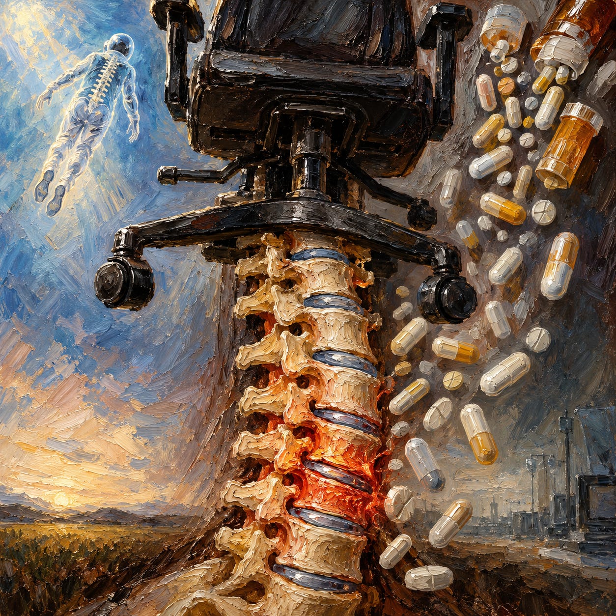
Author’s note: This essay treats height loss and the “osteoporosis” category as a case study in how a normal biological observation becomes a manufactured disease. When conventional terms like “osteoporosis,” “osteopenia,” “vitamin D,” “immune system,” and the various pharmaceutical class names appear, they carry the establishment’s own framing, which is the framing being prosecuted. My own analytical reading treats the body as a self-regulating organism responding to accumulated insult, and treats bone as a living, load-responsive tissue rather than a fixed genetic endowment.
You are one to two and a half centimeters shorter tonight than you were when you woke up.¹ This is not a metaphor. The intervertebral discs between your vertebrae are hydraulic structures with a tough outer ring and a gel-like inner core. They swell overnight when you lie horizontal and the compressive load of gravity is removed. They flatten again through the day as you stand and walk and sit. Every human being runs this experiment on themselves across every twenty-four-hour cycle. Height is not fixed.
The same mechanism, taken to the extreme, produces the two inches that NASA astronauts gain during six months on the International Space Station.² Once the constant downward push of gravity is removed, the spinal discs decompress across their full length. Scott Dulchavsky’s ultrasound imaging on the ISS has documented this process in real time, watching disc height and paraspinal muscle relaxation change over the course of a mission.³ Within months of returning to Earth, the astronaut shrinks back to preflight height.⁴ Loading conditions change; height responds.
Both observations demolish the story most doctors and most patients still accept about height loss with age. The story treats getting shorter as biological destiny, an inevitable part of the arc of aging that begins in the fifties and ends with a stooped grandparent. The actual evidence tells a different story. Spinal compression happens quickly. It reverses when the loading conditions change. It is directly tied to what the spine is being asked to carry. The variable is not calendar time. It is what is being done to the spine.
The two inches lost between age fifty and age seventy is the accumulated version of what a healthy body already undoes every twenty-four hours. What has to be explained is why the recovery stops happening. That question, followed properly, leads directly through the pharmacy and the office chair.
This work stays free because paid subscribers make it possible. They get the full book library, the Deep Dive Audio Library, and the Questions for Your Doctor, Before You Consent, and Package Insert series. No grants, no gatekeepers — your subscription is what keeps it that way.
The overnight test everyone runs
The intervertebral discs sit between each pair of vertebrae, roughly the size and shape of a hockey puck, made of a tough outer ring and a gel-like inner core with high water content. Under compressive load, fluid moves out. When compression is removed, fluid returns. The spine responds to loading conditions across many time scales, from the seconds of a squat to the decades of a life.
Adrian LeBlanc’s bed rest work at NASA in the 1980s established that patients placed at strict bed rest lose one to two percent of spinal bone mineral density per week.⁵ Bone resorption markers rise within the first days of a person going supine, and urinary calcium excretion rises on day one.⁶ The skeleton is not passively “aging.” It is reading the loading environment and adjusting accordingly. Remove the load, and it thins. Restore the load, and it rebuilds. This principle has a name in mainstream biomechanics, Harold Frost’s mechanostat, which holds that bone tissue continuously calibrates its density to the mechanical demands placed on it.
The astronaut and the bed-rested patient are the extreme cases of the same phenomenon that produces the shrinking grandparent. In each case, the spine is receiving instructions from what the body is doing all day. The grandparent’s instructions have been years of chair-sitting, minimal loading below ninety degrees of hip flexion, and often a growing list of prescriptions that interfere with bone remodeling directly. The bone and the discs are answering those instructions with the only response available to them.
The mechanical case
Patrick Daly, movement specialist at Tom Cowan’s New Biology Clinic, describes a pattern he sees across nearly every client who arrives complaining of back, hip, or knee pain. People stand and walk with their weight sitting back on their heels. Ask them to shift the weight forward, toward the ball of the foot, and they report the change feels foreign. The heel-loaded pattern has become their default. In Daly’s mechanical reading, this pattern locks the ankle joint, which is designed to be mobile, and forces the compensation upward through the chain: the knee, which is designed to be stable, is asked to move; the hip, designed to be mobile, clenches and tucks under; the lumbar spine flexes forward; the discs load unevenly and compress.⁷
The chain follows the established mobile-stable alternation that any physical therapy textbook will describe. Ankle mobile, knee stable, hip mobile, lumbar stable, thoracic mobile. When one link fails to do its job, the adjacent links absorb the demand and stiffen. Daly’s clinical observation, made over fifteen years of one-on-one work, is that most modern spinal complaints trace back down the chain to what the foot is doing. And what the foot is doing, in the population he sees, is riding the brake.
Chair-sitting adds another layer. Eight to ten hours a day in a chair loads the spine in flexion. The pelvis rolls into posterior tilt to accommodate the chair back. The hip flexors shorten and stay short. The Hadza hunter-gatherers of Tanzania spend roughly the same total hours per day sedentary as sedentary Americans do, but they rest by squatting or kneeling rather than by sitting in chairs.⁸ David Raichlen’s team at the University of Southern California measured muscle activity in both resting positions and found that squatting and kneeling maintain lower-limb muscle engagement that chair-sitting eliminates. The squat also loads the spine axially in the way the discs are built to accept. It keeps the hip through deep range. It keeps the ankle at full dorsiflexion. None of that is available in a chair.
Weston Price documented fourteen traditional populations across the globe in the 1920s and 1930s. Among them: the Loetschental Valley Swiss subsisting on rye bread and raw dairy from grass-fed cattle; the Gaelic islanders of the Outer Hebrides on oats and seafood; the Inuit of Alaska on seal, salmon, and organ meats; the Torres Strait Islanders on fish and coconut; the Maori of New Zealand, the Aboriginal Australians, the Melanesians, and the cattle-keeping East African tribes. The diets varied enormously. What Price observed did not. Dental arches were wide and dentition largely intact. The chronic conditions endemic to industrialized populations were essentially absent. Elderly members held upright posture into what Western observers would consider old age. The common factor across the groups was the absence of industrial processing in the food supply, combined with continual physical loading through daily work and movement.⁹
The mechanical case for why we shrink can be stated in plain terms. Modern life has removed the loading pattern that the spine and hip are built to respond to, and has replaced it with a loading pattern (chair-based, heel-weighted, static) that drives disc compression and postural collapse. Praveen Mummaneni, co-director of the UCSF Spine Center, has said openly that no systematic study has ever been funded to X-ray traditional populations and compare their spines to Western ones.¹⁰ That absence is itself informative. A finding that “sit on the floor and squat more” reduces the incidence of a condition that generates billions of dollars in scans, injections, surgeries, and drugs is not a finding anyone with money is eager to fund.
The pharmaceutical case
Mechanical compression alone would produce gradual height loss across a sedentary population. What has produced the modern acceleration, the fifty-year-old woman who is shrinking on the schedule her mother followed at seventy, is the pharmaceutical layer stacked on top. Multiple drug classes documented in mainstream literature to strip bone are now prescribed to substantial fractions of the adult population.
Corticosteroids are the most direct. Fitzpatrick’s review in the Mayo Clinic Proceedings series notes that patients on glucocorticoids can lose twenty to thirty percent of their trabecular bone within the first year of therapy.¹¹ Doses as low as 2.5 mg per day of prednisone are documented to produce bone loss. Fractures occur in thirty to fifty percent of long-term users. The medical literature calls this “glucocorticoid-induced osteoporosis,” a phrase that quietly admits the causal arrow: the drug produces the bone loss.¹² Corticosteroids are prescribed for a wide range of conditions labeled inflammatory or “autoimmune,” including asthma, rheumatoid arthritis, and skin conditions, and are given in short courses so routinely that most patients receiving them are not warned about the skeletal consequence.
Proton pump inhibitors were released with a heartburn indication and are now among the most prescribed drug classes in the world. Omeprazole (Prilosec), esomeprazole (Nexium), and lansoprazole (Prevacid) suppress the stomach’s production of acid. The FDA issued a Drug Safety Communication in May 2010 warning of increased risk of hip, wrist, and spine fractures with long-term or high-dose PPI use.¹³ The label warning followed. The proposed mechanism is that suppressing gastric acid impairs the conversion of dietary calcium salts into a form the intestine can absorb, producing chronic low-grade calcium malabsorption and, through it, elevated parathyroid hormone and increased bone resorption. Reviews in the gastroenterology literature have long noted that a substantial fraction of PPI prescriptions lack a clinical indication, with some estimates placing overprescription in the range of half or more of chronic users.¹⁴ Reflux typically responds to positional and dietary change; the drug does not fix the cause, and it steadily strips the bone.
Selective serotonin reuptake inhibitors were released as antidepressants and now account for roughly sixty percent of all antidepressant prescriptions in the United States. Wu and colleagues’ 2012 meta-analysis of thirteen cohort and case-control studies found SSRI users had a 72 percent higher fracture risk than non-users (RR 1.72).¹⁵ A larger 2020 meta-analysis by Kumar and colleagues that pooled 37 observational studies converged on a similar figure (RR 1.62).¹⁶ The mechanism appears to involve serotonin transporters on osteoblasts, the bone-building cells; blocking serotonin reuptake systemically disrupts bone formation. The UK Medicines and Healthcare products Regulatory Agency lists fracture risk in its official SSRI safety information.¹⁷ CDC survey data from 2019-2020 puts antidepressant use among US women at approximately 17 percent, with rates rising to 24 percent in women over sixty.¹⁸
The list continues. Aromatase inhibitors, prescribed to breast cancer patients, are documented to produce rapid bone loss. Loop diuretics, thyroid hormone over-replacement, anticonvulsants, and long-term heparin all appear in the medical literature as bone-stripping agents. Panday and colleagues’ 2014 review in Therapeutic Advances in Musculoskeletal Disease catalogs medication-induced osteoporosis across dozens of drug classes now in routine use.¹⁹ For the woman in her fifties on an SSRI, a PPI, occasional corticosteroids, and possibly thyroid replacement, the pharmaceutical assault on her skeleton is continuous.
The manufacture of a disease
The pharmaceutical story on bone loss did not begin with Fosamax. Through the 1970s and 1980s, hormone replacement therapy was prescribed to postmenopausal women partly on the argument that estrogen protected the bones. The Women’s Health Initiative trial, halted early in 2002 after documenting elevated rates of breast cancer and cardiovascular events in the HRT arm, ended that story.²⁰ HRT prescriptions dropped by approximately 39 percent within one year. Over the same period, prescriptions for the newer anti-osteoporosis medications, principally bisphosphonates, rose by approximately 29 percent as former HRT users were transitioned onto the newer bone drugs.²¹ Fosamax, which had been approved seven years earlier, moved into the space HRT vacated.
The story of how the pharmaceutical industry then turned the bone loss it was helping to cause into a formally marketable diagnosis is the establishment prosecuting itself. It began in June 1992 at a hotel near the Spanish Steps in Rome. The World Health Organization convened a small Study Group on osteoporosis, chaired by John Kanis, and asked it to produce a numerical definition of the condition.²² Merck, which had funded much of the development of dual-energy X-ray absorptiometry (DEXA) scanning technology, funded the meeting itself, as documented in Alix Spiegel’s 2009 NPR reporting on the wider marketing plan.²³ Before that meeting, osteoporosis had no numerical definition; doctors typically diagnosed it after a low-trauma fracture had already occurred.
The Study Group’s report, published in 1994 under the title Assessment of fracture risk and its application to screening for postmenopausal osteoporosis, introduced the T-score system.²⁴ A T-score compares a patient’s bone mineral density with the average density of a healthy thirty-year-old woman of the same race. The group set the cutoff for osteoporosis at 2.5 standard deviations below that reference (a T-score of -2.5), and introduced a new intermediate category called osteopenia for scores between -1.0 and -2.5. The choice was, in Kanis’s own later admission, arbitrary and never intended as a treatment threshold. Reviewing the history in a later paper, Karaguzel and Holick noted that the WHO cutoffs were designed as a prevalence measure and had since been repurposed, “as is common practice nowadays,” into a treatment trigger.²⁵
The consequence was immediate and predictable. Applying the criterion labeled roughly thirty percent of all postmenopausal Caucasian women as osteoporotic.²⁶ Adding the osteopenia category captured another fraction, so that approximately half of postmenopausal women now carried a bone-related label. In 1995, one year after the Kanis paper, the FDA approved Merck’s alendronate under the brand name Fosamax for the treatment of osteoporosis.
Merck’s marketing plan, described in detail by Alix Spiegel in her 2009 NPR report, then created the infrastructure required to convert the new diagnostic category into prescriptions.²⁷ Merck set up a nonprofit front called the Bone Measurement Institute, which had no payroll and no building. Its purpose was to promote the installation of DEXA scanners in doctors’ offices. Merck helped design and subsidize smaller, portable peripheral scanners that could sit on a desk. In 1995, there were roughly 750 bone-measuring devices in the United States. By 1999, there were between 8,000 and 10,000, according to Merck’s own count.²⁸ Merck lobbied Medicare to reimburse scans on the new machines, and Medicare bone-scan claims rose from 70,000 in 1994 to 1.5 million by 1999.
The circle then closed. Merck petitioned the FDA to approve a lower-dose version of Fosamax specifically for the treatment of osteopenia, the category that had been invented at the Rome meeting Merck had funded. The FDA approved it. The scan machines, now sitting in doctors’ offices, produced results as a traffic light: green for normal, yellow for osteopenia, red for osteoporosis. Women who arrived with a heartburn complaint or a menopausal question left with a bone scan, a yellow or red result, and a prescription.
John Kanis himself, the chairman of the 1992 WHO Study Group, has since confirmed in print that osteopenia was never intended as a disease category. It was a statistical designation for a portion of the reference distribution, and its conversion into a clinical diagnosis was, as Susan Kelleher documented at length in the Seattle Times in 2005, a marketing achievement rather than a medical one.²⁹
The final layer of the paradox
The bisphosphonate class, of which Fosamax was the first, works by killing osteoclasts, the cells that break down old bone in the normal remodeling cycle. On a bone scan, the effect looks like preserved density. Underneath, the bone is not being remodeled. Old, brittle bone accumulates because the cellular machinery for replacing it has been chemically suppressed.
The DEXA scan itself is part of the deception. The scanner measures bone mineral density, which is how much mineral is packed into a given volume. It does not measure bone quality. It does not measure microarchitecture or resilience, and it does not measure whether the bone can absorb stress without shattering. Bisphosphonate-treated bones score higher on DEXA precisely because the drug suppresses the remodeling that would remove and replace old, brittle tissue. The old tissue accumulates. On the scan the density looks improved. Under mechanical load, that same bone fails at forces a normally remodeling skeleton would absorb without incident.
The measurement problem shows up in the fracture data. Siris and colleagues, drawing on the National Osteoporosis Risk Assessment cohort of 149,524 postmenopausal women, reported that only 18 percent of women who sustained an osteoporotic fracture in the year following their DEXA scan had a T-score of -2.5 or below.³⁰ Eighty-two percent of the women who actually fractured would not have qualified for osteoporosis treatment under the WHO’s own threshold. The category the entire diagnostic and pharmaceutical infrastructure was built around fails as a fracture predictor in the population it is applied to. The measurement rewards the drug that suppresses remodeling, and misses the women who will actually break.
Ten years into the Fosamax era, the consequence appeared in emergency rooms. Long-term bisphosphonate users began arriving with atypical femur fractures, breaks in the shaft of the thigh bone, occurring under trivial loads or spontaneously while standing. In October 2010, the FDA added a warning to the labels of all bisphosphonates about the increased risk of atypical subtrochanteric and diaphyseal femur fractures.³¹ An American Society for Bone and Mineral Research task force reviewed 310 atypical femur fracture cases and found that 94 percent of the patients had been taking bisphosphonates, most for more than five years.³² Osteonecrosis of the jaw, in which the jawbone dies and sloughs off, was added to the labels as another rare but documented consequence.
Defenders of the drug class point, correctly, to trial data showing that in the first three to five years of use, in women who have genuine osteoporosis and have already sustained a low-trauma fracture, bisphosphonates reduce vertebral fracture rates and in some studies hip fracture rates. This is real. It is also not what the drug is being prescribed for in most cases. The osteopenia category invented at the 1992 Rome meeting captured women who did not have osteoporosis and had never fractured. Merck’s low-dose Fosamax was approved specifically for that population. And prescriptions have never been limited to three or five years. Women are put on bisphosphonates in their fifties, told to stay on them indefinitely, and reach year seven or eight or ten with no plan to stop. The window in which the drug provides fracture protection closes well before the window in which the atypical fractures begin.
The most consequential finding from the FDA’s own long-term extension data has received less coverage. In a pooled analysis of the three major bisphosphonate extension studies, patients who continued the drug beyond six years had non-vertebral fracture rates of 9.3 to 10.6 percent, versus 8.0 to 8.8 percent for those switched to placebo.³³ Continuing bisphosphonate treatment beyond five years produced no additional fracture protection and added the atypical fracture risk. The drug that was sold to prevent fractures, taken long enough, produces new kinds of fractures without reducing the total.
The pharmaceutical industry created the conditions (bone-stripping side effects across the top-prescribed drug classes), sat on the panel that invented the diagnostic category (osteopenia was defined at a meeting Merck funded), placed the scanning equipment in the offices (via a nonprofit shell it created), and sold the drugs that, over the durations they were actually prescribed for, produced new fractures without reducing overall fracture rates. This is not conspiracy theorizing. It is a chronology assembled from the FDA’s own actions, the WHO’s own committee report, Merck’s own admissions in the NPR reporting, and mainstream orthopedic literature.
Why the modern epidemic is worse
The two pillars, mechanical and pharmaceutical, are not independent. They compound. A woman born in 1975 has been in classroom chairs since age five, moved to a desk chair through university, and settled into an office chair through her working years. She has essentially never squatted below ninety degrees of hip flexion since childhood. Her postural default is heel-weighted and forward-headed. On top of that mechanical substrate, by her mid-fifties she is statistically likely to be on some combination of the drugs listed above: an SSRI or SNRI, a PPI, possibly a thyroid replacement, occasional corticosteroid courses for skin or joint or respiratory conditions, and, once her first DEXA scan comes back yellow or red, a bisphosphonate.
Her mother, born in 1935, had none of that pharmacology available for most of her adult life. She spent decades doing housework that involved kneeling, squatting, bending, reaching, and carrying. She had children in an era when floor-level care of small children was still the norm. Her diet included more whole animal foods and less industrial oil, though it too had been degraded by the post-war shift. She held her height into her seventies, and lost perhaps an inch by her mid-eighties.
The comparison holds beyond nostalgia. One spine has been subjected to two orthogonal insults running for fifty years. The other spine had substantially less of either. The mother’s tissue was under the loading pattern it was designed for, without pharmaceutical interference with the remodeling cycle. The daughter’s tissue has been running the opposite protocol continuously since she was five years old.
Four converging lines on reversal
Height loss is not one-way. The evidence for reversal comes from four independent directions, and their convergence is what makes the case difficult to dismiss.
The first line is the astronaut return-to-Earth data. Dulchavsky’s ongoing ultrasound work at NASA has documented that the height gained in microgravity returns to preflight baseline within months of landing, and that bone density recovers partially, faster after longer flights. Stavnichuk and colleagues’ 2020 systematic review of the aerospace bone-loss literature found that in the months following return to Earth, bone formation markers rise linearly at 84 percent per month above pre-flight levels for the first three to five months.³⁴ The mechanism is exactly what would be predicted from Frost’s mechanostat: restore the loading, the tissue rebuilds. Bone is not a static substance. It is a continuously remodeling tissue reading its environment.
The second line comes from bed rest reversal studies. The same laboratories that documented rapid loss with immobilization documented that once the subjects resumed ambulation and weight-bearing, bone markers and density began to recover on a symmetric timeline.³⁵ The stimulus that caused the loss, when reintroduced, caused the recovery.
The third line is pharmaceutical discontinuation. Kim and colleagues’ Korean nationwide cohort found that fracture risk in former SSRI users declines toward baseline after cessation.³⁶ Post-corticosteroid recovery data has been reviewed in the Cleveland Clinic Journal of Medicine: bone loss following glucocorticoid discontinuation is partially reversible, with the majority of recovery occurring in the first year off the drug.³⁷ Getting off the drug that was stripping the bone is a real intervention with documented effect. It does not require a new drug to reverse the damage of the old one, though the entire industry is now organized around selling exactly that.
The fourth line is cross-cultural. Traditional populations that continue to squat, kneel, and load the spine axially throughout life show minimal age-related height loss. This is observational evidence, and as Mummaneni acknowledges, no systematic imaging study has been funded to characterize their spines. The absence of that funding is itself a data point. A finding that would translate to “squat more, sit less, and get off the drugs” generates no prescriptions, and so it does not get investigated.
Patrick Daly’s clinical observation completes the picture at the practical level. His fifteen years of one-on-one work at New Biology Clinic have accumulated many cases of clients who have regained measurable height and reversed years of postural collapse. The interventions are unglamorous. The weight comes off the heel and back through the foot. Gentle axial loading gets added through squats and deep hip positions. The person slows down enough to feel where the body has been bracing, and the bracing releases. This is not trial-grade evidence. It does not need to be. The mechanism has already been documented at the extremes by NASA and by immobilization studies. Daly’s work shows what the mechanism looks like at ordinary scale, in ordinary bodies.
Closing
The two inches lost by age seventy is not the arithmetic of biology. It is the accumulated cost of continuous compression stacked on top of a pharmaceutical layer that interferes with bone remodeling. On top of both sits a diagnostic category, invented at a meeting funded by the company that sold the drug for it. Every one of those variables sits inside the range of choice.
The astronaut who returns from six months on the ISS and regains lost height within months of landing is not exhibiting an exotic phenomenon. It is what a functioning spine does when the load path is restored. Every human body runs a smaller version of the same experiment across every twenty-four-hour cycle, gaining and losing a centimeter or two between waking and sleeping. What has been sold as inevitable was in fact assembled. The chair did some of it. The prescription did more of it. A WHO committee sitting in a Rome hotel room in 1992, with Merck picking up the tab, provided the diagnostic frame that made the drug prescribable.
The question of why we shrink turns out to be the question of why the recovery stops. The answer sits mostly in the office chair and mostly in the medicine cabinet. Both are within reach.
How to Explain It to a Six-Year-Old
Your back is a stack of small bones with soft cushions between them. The cushions are like little water balloons. When you lie down at night, the balloons fill up with water. When you stand up all day, the water squishes out a bit. That’s why you’re a little taller in the morning than at bedtime.
If you sit slumped in a chair for many hours every day, and never sit on the floor or squat down like a frog, the cushions get thinner over time and stop filling up all the way. If you also take medicines that make the cushions get thinner, the cushions get even thinner even faster. This is why some grownups get shorter as they get older. It isn’t because their body is broken. It’s because of how they sit and what they take.
Astronauts float in space and don’t have gravity pushing down on them. Their cushions fill up so much they get two inches taller. When they come back to Earth, they get short again. That shows the cushions are alive and changing all the time. If you squat like a frog, and don’t sit slumped for hours, and don’t take medicines that squish your cushions, your cushions stay full and plump for a very long time. Getting shorter isn’t something that just happens. It’s something the body does when we do certain things to it.
In Print
The Unbekoming library is available in paperback, printed to order through Lulu and shipped worldwide. The shelf begins with the paradigm question underneath everything else — No Virus, the isolation problem, the collapse of virology’s foundational claims, and a disease-by-disease reappraisal — and moves through the suppressed compounds mainstream medicine set aside: The DMSO Book, Chlorine Dioxide: The Forbidden Remedy, The Iodine Book, and The Hydrogen Peroxide Book. Two more recover what’s still on the kitchen shelf: Baking Soda and The Castor Oil Book. Two more recover the minerals modern soil, water, and processing quietly stripped from the diet: The Magnesium Handbook and The Boron Book. Sitting alongside these is No Contagion, co-authored with Jamie Andrews — the case against germ theory itself, catalogued through 258 failed contagion experiments.
The critique books cover what medicine, dentistry, psychiatry, and veterinary practice have become. The Unvaccinated treats the completely unvaccinated as a comparison group across twenty chapters and five appendices. Medicalized Motherhood follows a woman through 123 documented interventions from teenage pill to postpartum discharge. Drilling for Profit treats cavities, gum disease, and crooked teeth as the dietary problem they are. What Your Vet Can’t Tell You applies the same critique to pets. Escape from Psychiatry documents the fabrication of the DSM and the specific damage of every major psychiatric drug class. The Vitamin K Injection covers what happens in the first hours of a newborn’s life.
The full shelf is at lulu.com/spotlight/unbekoming. A physical book reaches the person a Substack post never will — the skeptical relative, the friend who won’t click a link but might open a book, the visitor whose eye lands on a coffee table. Buy one to keep, and one to give away.
This essay is written for educational and analytical purposes. It is not medical advice. Anyone considering changes to prescribed medications should consult a physician who is willing to engage seriously with the questions raised here.
New Biology Clinic
For those of you looking for practitioners who actually understand terrain medicine and the principles we explore here, I want to share something valuable. Dr. Tom Cowan—whose books and podcasts have shaped much of my own thinking about health—has created the New Biology Clinic, a virtual practice staffed by wellness specialists who operate from the same foundational understanding. This isn’t about symptom suppression or the conventional model. It’s about personalized guidance rooted in how living systems actually work. The clinic offers individual and family memberships that include not just private consults, but group sessions covering movement, nutrition, breathwork, biofield tuning, and more. Everything is virtual, making it accessible wherever you are. If you’ve been searching for practitioners who won’t look at you blankly when you mention structured water or the importance of the extracellular matrix, this is worth exploring. Use discount code “Unbekoming” to get $100 off the member activation fee. You can learn more and sign up at newbiologyclinic.com
References
Reilly T, Tyrrell A, Troup JD. “Circadian variation in human stature.” Chronobiology International 1, no. 2 (1984): 121–126.
Wall M. “Strange but True: Astronauts Get Taller in Space.” Space.com, January 7, 2013. See also NASA, “Space Bones,” NASA Science News, October 2001.
Sayson JV, Lotz J, Parazynski S, Hargens AR. “Back pain in space and post-flight spine injury: mechanisms and countermeasure development.” Acta Astronautica 86 (2013): 24–38. See also Dulchavsky S, principal investigator, ISS spinal ultrasound study, NASA.
Adventist Health System. “How Astronaut Scott Kelly Got Two Inches Taller While in Space.” AdventHealth Blog.
LeBlanc A, Schneider V, Krebs J, Evans H, Jhingran S, Johnson P. “Spinal bone mineral after 5 weeks of bed rest.” Calcified Tissue International 41 (1987): 259–261.
Baecker N, Tomic A, Mika C, et al. “Bone resorption is induced on the second day of bed rest: results of a controlled crossover trial.” Journal of Applied Physiology 95, no. 3 (2003): 977–982.
Daly P. “Healing Is Hell When You’re Heavy on Your Heels.” New Biology Clinic webinar, August 26, 2026. Transcript on file.
Raichlen DA, Pontzer H, Zderic TW, et al. “Sitting, squatting, and the evolutionary biology of human inactivity.” Proceedings of the National Academy of Sciences 117, no. 13 (2020): 7115–7121.
Price WA. Nutrition and Physical Degeneration. 8th ed. La Mesa, CA: Price-Pottenger Nutrition Foundation, 2008 (original 1939).
Mummaneni P, interviewed by Michaeleen Doucleff. “Lost Posture: Why Some Indigenous Cultures May Not Have Back Pain.” NPR Morning Edition, June 8, 2015.
Fitzpatrick LA. “Secondary causes of osteoporosis.” Mayo Clinic Proceedings 77, no. 5 (2002): 453–468.
Buckley L, Humphrey MB. “Glucocorticoid-Induced Osteoporosis.” New England Journal of Medicine 379 (2018): 2547–2556. See also American College of Rheumatology 2017 Guideline for the Prevention and Treatment of Glucocorticoid-Induced Osteoporosis.
US Food and Drug Administration. “FDA Drug Safety Communication: Possible increased risk of fractures of the hip, wrist, and spine with the use of proton pump inhibitors.” May 25, 2010.
Ito T, Jensen RT. “Association of long-term proton pump inhibitor therapy with bone fractures and effects on absorption of calcium, vitamin B12, iron and magnesium.” Current Gastroenterology Reports 12, no. 6 (2010): 448–457. See also Forgacs I, Loganayagam A, “Overprescribing proton pump inhibitors,” BMJ 336, no. 7634 (2008): 2–3.
Wu Q, Bencaz AF, Hentz JG, Crowell MD. “Selective serotonin reuptake inhibitor treatment and risk of fractures: a meta-analysis of cohort and case-control studies.” Osteoporosis International 23, no. 1 (2012): 365–375.
Kumar M, Bajpai R, Shaik AR, et al. “Alliance between selective serotonin reuptake inhibitors and fracture risk: an updated systematic review and meta-analysis.” European Journal of Clinical Pharmacology 76, no. 10 (2020): 1373–1392.
UK Medicines and Healthcare products Regulatory Agency. “Selective Serotonin Reuptake Inhibitors (SSRI), Learning Module, Section 4.13: Bone fractures.” MHRA Continuing Professional Development.
Brody DJ, Gu Q. “Antidepressant Use Among Adults: United States, 2015–2018.” NCHS Data Brief No. 377, September 2020. Centers for Disease Control and Prevention, National Center for Health Statistics.
Panday K, Gona A, Humphrey MB. “Medication-induced osteoporosis: screening and treatment strategies.” Therapeutic Advances in Musculoskeletal Disease 6, no. 5 (2014): 185–202.
Writing Group for the Women’s Health Initiative Investigators. “Risks and benefits of estrogen plus progestin in healthy postmenopausal women: principal results from the Women’s Health Initiative randomized controlled trial.” JAMA 288, no. 3 (2002): 321–333.
Farley JF, Blalock SJ, Cline RR. “Effect of the Women’s Health Initiative on prescription anti-osteoporosis medication utilization.” Osteoporosis International 19, no. 11 (2008): 1603–1612.
Kanis JA. “Assessment of fracture risk and its application to screening for postmenopausal osteoporosis: synopsis of a WHO report. WHO Study Group.” Osteoporosis International 4, no. 6 (1994): 368–381.
Yoho R. “Osteoporosis: How Pharma Invented a Fake Diagnosis, Sold Poison Drugs, and Hid the Real Cures.” Dr Yoho podcast and essay 460, July 22, 2026. See also Spiegel A, “How a Bone Disease Grew to Fit the Prescription,” NPR All Things Considered, December 21, 2009.
World Health Organization. Assessment of fracture risk and its application to screening for postmenopausal osteoporosis. WHO Technical Report Series 843. Geneva: WHO, 1994.
Karaguzel G, Holick MF. “Diagnosis and treatment of osteopenia.” Reviews in Endocrine and Metabolic Disorders 11 (2010): 237–251.
Kanis JA, Melton LJ, Christiansen C, Johnston CC, Khaltaev N. “The diagnosis of osteoporosis.” Journal of Bone and Mineral Research 9, no. 8 (1994): 1137–1141.
Spiegel A. “How a Bone Disease Grew to Fit the Prescription.” NPR All Things Considered, December 21, 2009.
SourceWatch. “Bone Measurement Institute.” Center for Media and Democracy. Documents Merck’s creation of the Bone Measurement Institute and the growth of DEXA scanner deployment from 750 in 1995 to between 8,000 and 10,000 by 1999.
Kelleher S. “Disease expands through marriage of marketing and machines.” Seattle Times, June 28, 2005.
Siris ES, Chen YT, Abbott TA, et al. “Bone mineral density thresholds for pharmacological intervention to prevent fractures.” Archives of Internal Medicine 164, no. 10 (2004): 1108–1112. Data drawn from the National Osteoporosis Risk Assessment (NORA) study cohort of 149,524 postmenopausal women.
US Food and Drug Administration. “FDA Drug Safety Communication: Safety update for osteoporosis drugs, bisphosphonates, and atypical fractures.” October 13, 2010.
Shane E, Burr D, Ebeling PR, et al. “Atypical subtrochanteric and diaphyseal femoral fractures: report of a task force of the American Society for Bone and Mineral Research.” Journal of Bone and Mineral Research 25, no. 11 (2010): 2267–2294.
Black DM, Kelly MP, Genant HK, et al. “Bisphosphonates and fractures of the subtrochanteric or diaphyseal femur.” New England Journal of Medicine 362, no. 19 (2010): 1761–1771. See also FDA background material for the September 2011 Advisory Committee meeting on long-term bisphosphonate use.
Stavnichuk M, Mikolajewicz N, Corlett T, Morris M, Komarova SV. “A systematic review and meta-analysis of bone loss in space travelers.” npj Microgravity 6 (2020): 13.
Rittweger J, Frost HM, Schiessl H, et al. “Muscle atrophy and bone loss after 90 days’ bed rest and the effects of flywheel resistive exercise and pamidronate: results from the LTBR study.” Bone 36, no. 6 (2005): 1019–1029.
Kim JH, et al. “Use of serotonin reuptake inhibitors and risk of subsequent bone loss in a nationwide population-based cohort study.” Scientific Reports 11 (2021): 13461.
Cleveland Clinic Journal of Medicine. “Glucocorticoid-induced osteoporosis: Insights for the clinician.” CCJM 87, no. 7 (2020): 417–426.
Additional Sources
Cowan T. Human Heart, Cosmic Heart. White River Junction, VT: Chelsea Green, 2016.
Shelton HM. Natural Hygiene: Man’s Pristine Way of Life. Available through Natural Hygiene Press.
Tilden JH. Toxemia Explained: The True Interpretation of the Cause of Disease. 1926.
Firstenberg A. The Invisible Rainbow: A History of Electricity and Life. White River Junction, VT: Chelsea Green, 2020.
Purdey M. Animal Pharm. East Sussex: Clairview Books, 2007.
Latypova S. Due Diligence and Art. 2024.
Williams C, Bailey P. Terrain Therapy. Self-published, 2022.
My man! Here’s a supplementary article to this one. I recently wrote about why we need to remain athletic — even into our 50s, 60s, and 70s — and it ties perfectly into what you’ve written here.
We can and should continue getting stronger as we age, even though modern society seems designed to steal our strength and vitality away from us.
Very timely piece. Here’s my article on aging and remaining athletic:
https://unorthodoxy.substack.com/p/why-you-should-be-athletic-at-any
Where can I leave a question and comment on your new book I just received… The Magnesium Handbook???