

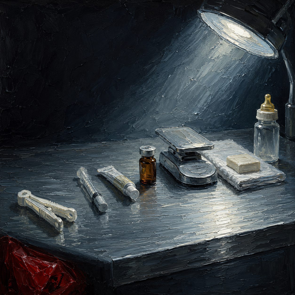
In the first three minutes after the infant is delivered, if the umbilical cord is left intact, approximately one hundred milliliters of blood transfers from the placenta into the newborn.¹ That is roughly a third of the infant’s total blood volume. The transfer is not passive. The umbilical vessels contain their own smooth muscle. They close on their own schedule when the transfer is complete, usually within three to ten minutes. At that point the cord blanches, the pulsation stops, and the vessels seal.
In the United States, this process is interrupted, on average, within thirty to sixty seconds. The infant loses roughly thirty percent of the blood the physiology was constructed to deliver. The practice is defended on grounds that shift as the literature closes on it: it prevents jaundice (it does not, at any clinically meaningful rate), it reduces polycythemia (rarely, and asymptomatically), it is what the obstetrician was trained to do (this last is accurate). The iron the infant would have used for the next six months goes into a red plastic bag with the placenta.
This essay is about what happens in the first sixty minutes after birth in a standard American hospital, and about the biology of the first sixty minutes as it existed before the interventions displaced it. There are seven interventions in the standard sequence. Each one displaces a specific biological event. The essay traces the seven in the order they occur, and then examines the second casualty of the hour, which is the mother.
The argument is not that any single intervention is catastrophic in isolation. It is that the biology of the first hour was doing something, and hospital protocol displaces the entire sequence in favor of procedures whose cumulative effect has never been studied. What the interventions replace is a sequence humans arrived with. What replaced it was assembled between roughly 1930 and 1991, most of it introduced without controlled trial, and none of it evaluated as the sequence it now is.
The infant born in a Kansas farmhouse in 1890 received the first hour by default. The infant born in a Manhattan delivery room in 2026 receives the first hour only if the parents have prepared for months to protect it against interventions the hospital considers routine.
This work stays free because paid subscribers make it possible. They get the full book library, the Deep Dive Audio Library, and the Questions for Your Doctor, Before You Consent, and Package Insert series. No grants, no gatekeepers — your subscription is what keeps it that way.
Audio Deep Dive (for Paid Subscribers)
The Placental Transfer
The Cochrane review on the timing of umbilical cord clamping in term infants pools twenty-five trials involving more than three thousand mother-infant pairs. Delayed clamping (variously defined as one to three minutes, or until pulsation ceases) is associated with higher hemoglobin at birth, higher ferritin at three and six months, and lower rates of iron deficiency in infancy.² The World Health Organization recommends delaying cord clamping for at least one minute after birth.³ The American College of Obstetricians and Gynecologists, in Committee Opinion Number 814 (January 2020), endorsed a delay of at least thirty to sixty seconds. Actual hospital practice in the United States has moved reluctantly, and inconsistently, toward the low end of that range.
Ola Andersson and colleagues at Uppsala University randomized four hundred term infants to early clamping (within ten seconds) or delayed clamping (at least three minutes) and followed them at four months and at four years. At four months, the delayed-clamping infants had ferritin levels forty-five percent higher than the early-clamping group.⁴ At four years, the delayed-clamping children scored higher on fine motor and social skills.⁵ The trial was published in the BMJ and in JAMA Pediatrics. It has not changed what happens in the room where the baby comes out.
The infant delivered at term arrives with approximately eighty milliliters of blood per kilogram in circulation. The placenta at the moment of birth contains roughly one third of the total fetal blood volume. That fraction is not surplus. The umbilical arteries and vein complete the redistribution while the infant’s lungs open and the ductus arteriosus and foramen ovale begin the transition from fetal to neonatal circulation. The iron load carried in the umbilical vessels at three minutes postpartum is approximately 70 milligrams. The daily iron requirement of the term infant during the first six months, met almost entirely from stores laid down before and during the third trimester, is approximately 0.27 milligrams. The math is not subtle. Three minutes of transfer delivers nine months of iron.
The American replacement is a plastic clamp on a still-pulsing cord, and a pair of scissors thirty seconds later. The consequences are measurable at four months (ferritin, hemoglobin), at six months (iron sufficiency), and at four years (fine motor and social scoring). The intervention has no informed consent process. The mother will rarely be told that the WHO and ACOG recommendations both call for a longer delay than the hospital in fact provides, and she will almost never be told what the delayed-clamping trials measured at four years.
The Approach to the Breast
The infant placed on the mother’s abdomen immediately after birth, undisturbed, dry but not washed, performs a sequence of movements that has been documented on video and named. Ann-Marie Widström and colleagues in Sweden videotaped twenty-eight undisturbed term newborns and identified nine sequential stages: birth cry, relaxation, awakening, activity, crawling, resting, familiarization, suckling, and sleeping.⁶ The sequence completes, on average, in the first sixty to ninety minutes after birth. The infant uses head-lifting movements, hand-mouth coordination with the mother’s nipple, and a slow crawl up the mother’s abdomen to the breast. The first latch, when the infant is left to accomplish it, typically occurs somewhere between sixty and ninety minutes after birth, with substantial variation between infants.
The Cochrane systematic review on early skin-to-skin contact between mother and healthy newborn pools forty-six trials with more than three thousand mother-infant pairs. Infants who received skin-to-skin contact in the first hour, without separation for measurement or procedure, breastfed earlier, breastfed longer, maintained more stable blood glucose, thermoregulated more efficiently, and cried less in the following twenty-four hours.⁷ Maternal outcomes included lower rates of breastfeeding cessation before six weeks and lower postpartum depression scores.
Standard American hospital practice moves the infant to a warmer within the first minute. There the infant is weighed, measured, footprinted, given an Apgar score, injected with the compound sold as vitamin K, and treated with erythromycin eye ointment. The infant is then wrapped in a receiving blanket and returned to the mother. The nine-stage sequence has been interrupted at stage three. The rest of the stages do not resume. The first latch, if it occurs in the first hour at all, occurs against the mother’s blanket-wrapped chest, with an infant whose hand-mouth coordination has been disrupted, whose skin has been rubbed with an absorbent towel, and whose eyes have been coated with antibiotic ointment.
What is displaced in the assessment ritual is the imprinting window. The infant returned to the mother wrapped, cleaned, and processed does not get the first hour. Neither does the mother.
The First Injection
The compound sold as vitamin K is administered to the American newborn by intramuscular injection, typically in the thigh, within the first fifteen minutes of life. The standard dose is one milligram. Endogenous plasma phylloquinone in the term newborn is approximately 0.05 nanograms per milliliter. The injection delivers a bolus at roughly twenty thousand times endogenous. The formulation includes polysorbate 80, benzyl alcohol, and propylene glycol.⁸ The active compound, phytonadione, is a synthetic form of phylloquinone.
The intervention has a real underlying observation. What mainstream pediatrics calls vitamin K-dependent bleeding, formerly labeled hemorrhagic disease of the newborn, occurs in a small fraction of untreated infants during the first week of life and, in a smaller fraction, between two weeks and six months. The late-onset form can produce intracranial hemorrhage and carries substantial mortality when it occurs. The bleeding phenomenon is not disputed. What is disputed is what to do about it.
The American Academy of Pediatrics adopted routine intramuscular prophylaxis in 1961.⁹ The route was chosen for the assurance of absorption, not because oral phytonadione does not work. Multiple oral regimens have been tested and are used in national protocols in the Netherlands, Denmark, Switzerland, and Germany. The Dutch protocol delivers 1 milligram at birth followed by 150 micrograms daily from day eight to week thirteen in breastfed infants. Rates of late-onset bleeding in the Dutch protocol are comparable to the American intramuscular protocol.¹⁰
Jean Golding and colleagues at Bristol published in the British Medical Journal in 1992 an analysis linking the intramuscular injection at birth to a doubling of childhood cancer, particularly leukemia, in the exposed cohort.¹¹ Subsequent studies were mixed. The mainstream position settled into “the balance of evidence does not support the association,” and the intramuscular protocol continued. The oral protocol, which delivers the same effect at physiologic dosing without the injection additives, was not adopted in the United States.
The additives merit examination on their own. Benzyl alcohol at neonatal doses has been associated with gasping syndrome in premature infants; the FDA issued a warning in 1982 restricting its use in that population.¹² The one-milligram vitamin K injection contains 9 milligrams of benzyl alcohol. Polysorbate 80 is a solubilizer with documented capacity to disrupt cellular membrane integrity and, in animal models, alter blood-brain barrier permeability. A prior essay developed the zeta-potential mechanism at length.¹³ The vitamin K injection is not the largest zeta-potential insult of the first hour. It is the first.
What the injection displaces is the intended pattern of vitamin K acquisition, which is neither zero nor bolus. The breastfed infant receives phylloquinone in milk at physiologic concentrations sustained across the first months. The intramuscular protocol delivers, in a single dose, an amount the physiology was constructed to receive across weeks.
For the reader wondering whether their own child received this injection: consent was almost certainly not obtained in any meaningful sense. A form signed on admission authorized the hospital’s standard newborn care. That form covered the injection.
The Ointment
Within the first thirty minutes, before the infant is returned to the mother, erythromycin ophthalmic ointment (0.5 percent) is applied to both eyes. The procedure is required by law in most states. The rationale, dating to Karl Credé’s 1881 protocol in Leipzig, was prevention of what mainstream medicine calls ophthalmia neonatorum, a neonatal eye inflammation the establishment attributes to gonococcal exposure during birth. Untreated, the condition was documented to progress to corneal ulceration and blindness in a subset of affected infants. Credé’s silver nitrate reduced the reported incidence in Leipzig from ten percent of newborns to less than one percent. The intervention reduced blindness. The intervention that replaced it, applied to a screened-negative population, does not.
Silver nitrate has since been replaced by erythromycin. The current protocol treats the infant of a screened mother not carrying the bacterium, in a population where gonorrhea prenatal screening is standard care.¹⁴ ACOG recommends screening at the first prenatal visit for all pregnant patients under twenty-five and for older patients with risk factors. A mother screened and treated presents what the establishment characterizes as no meaningful risk of transferring the bacterium. The ointment is applied anyway. The state mandate does not distinguish between screened-negative and unscreened mothers.
The ointment blurs the infant’s vision during the imprinting window. The newborn is optically calibrated to focus at eight to twelve inches, the distance from breast to mother’s face during nursing. Ointment-coated eyes cannot fix on that face. The ointment is also an antibiotic administered to the ocular surface at the moment the ocular microbiome is being established. No trial has ever measured what the ointment does to that colonization. The absence of the trial is the finding.
The eye ointment is one of the interventions parents can decline in most jurisdictions with a signed refusal form. It is rarely offered as an option. The nurse arriving with the tube does not typically pause to ask.
The Vaccination
At some point in the first twenty-four hours, and in some hospital protocols within the first hour or two, the newborn receives the first injection on the American vaccination schedule. The vaccine is the recombinant hepatitis B vaccine, marketed as Recombivax HB or Engerix-B, containing 5 to 10 micrograms of hepatitis B surface antigen and 250 micrograms of aluminum, as amorphous aluminum hydroxyphosphate sulfate (Recombivax) or aluminum hydroxide (Engerix-B).¹⁵
The rationale for administering this injection on the day of birth was established by the ACIP in 1991 as part of a strategy to eliminate what mainstream medicine calls hepatitis B transmission through universal infant vaccination.¹⁶ In the establishment framework, the condition is associated with blood-to-blood contact and sexual activity. The pediatric concern the ACIP named was vertical transfer from a mother carrying the surface antigen. Screening for that surface antigen has been standard prenatal care since 1988. The mother screened and confirmed negative delivers an infant whose risk of what the establishment calls hepatitis B in the first year of life, absent blood contact and sexual activity, approaches zero on the establishment’s own terms. The rationale for injecting that infant on the day of birth reduces to catching the infants of unscreened mothers, and to protecting against later transfer from a household member sharing razors. Both are surrogate arguments for injecting a screened-negative population.
The infant on day one receives 250 micrograms of aluminum in a single bolus. The FDA has identified 4 to 5 micrograms per kilogram per day as the exposure level at which parenteral aluminum accumulates to concentrations associated with central nervous system and bone toxicity in patients with impaired renal function.¹⁷ Applied to a 3.5-kilogram newborn, the FDA’s accumulation threshold works out to roughly 17 micrograms per day. The day-one injection delivers about fifteen times that amount, in a single bolus, into an infant whose kidneys are not yet fully working and whose blood-brain barrier will never again be this porous.
The aluminum-adjuvant literature, developed most extensively by Christopher Exley at Keele and Romain Gherardi at Créteil, documents that injected aluminum does not remain at the site. Macrophages carry it to lymph nodes, spleen, bone marrow, and, in Gherardi’s macrophagic myofasciitis series, to distant tissue including brain.¹⁸ ¹⁹ Clearance from tissue, once deposited, is measured in years. Zeta potential collapse in the neonatal blood following aluminum-adjuvant injection is the mechanism a separate essay in this series developed. The injection arrives when the infant is least prepared to receive it. The schedule is calibrated to that moment on purpose.
The 1986 National Childhood Vaccine Injury Act removed vaccine manufacturer liability for injuries arising from the recommended pediatric schedule.²⁰ A prior essay in this series traced the consequences of that legal structure. The injection administered to the newborn is the first of the schedule the Act protects. Consent is bundled into admission paperwork. The parent who declines is required to sign a separate refusal form. The counseling does not mention the aluminum load, the biodistribution literature, or the mother’s own negative screening.
The Vernix
The white coating on the newborn’s skin is not residue. It is a substance the infant made, for the infant, starting in the second trimester of gestation, composed of eighty percent water, ten percent protein, and ten percent lipid. The protein fraction contains at least forty-one distinct proteins identified in the Tollin proteomic analysis, of which roughly a third have direct antimicrobial activity.²¹ The active components include lysozyme, lactoferrin, cathelicidin LL-37, and a range of defensins active against gram-positive and gram-negative bacteria, fungi, and some enveloped particles. The lipid fraction, in composition and quantity, closely tracks the composition of the stratum corneum (the outermost skin layer) of the term infant. It is not a coincidence.
The vernix serves at least four documented functions. In utero it prevents maceration of the fetal skin by amniotic fluid across the third trimester. At delivery it eases passage through the birth canal. Post-delivery it retains warmth and moisture at the skin surface, providing thermoregulation and preventing transepidermal water loss. And it colonizes the infant’s skin with an antimicrobial coating whose peptides continue functioning during the hours in which the skin microbiome is being established.
The World Health Organization recommends delaying the first bath by at least twenty-four hours after birth.²² American hospital practice varies. Many hospitals wash the infant within the first several hours, sometimes with soap. The vernix is rubbed off with towels during drying and the residue is washed away in the bath.
What the vernix does that no substitute is offered for is the seeding of the infant’s skin surface with the mother’s microbiome combined with the fetal-secretion antimicrobial peptides. The infant delivered vaginally, placed on the mother’s chest unwashed, receives the mother’s flora onto skin coated with the vernix, which selects for the flora the peptides tolerate and against the flora the peptides suppress. The infant washed, wrapped in hospital linen, and handled by gloved hands receives hospital flora onto skin whose antimicrobial coating has been removed.
Maria Dominguez-Bello and colleagues have documented that the microbiome of the cesarean infant at one month differs measurably from the vaginally-delivered infant at one month, with the cesarean infant’s early flora more closely resembling adult skin flora than the mother’s vaginal flora.³⁰ Subsequent work has shown that at least some of the differences persist through infancy.
The vernix is not a candidate for pharmaceutical replacement. There is no product to sell in its place. It was produced by the infant, for the infant, and it functions in a window that closes within hours. What replaces the vernix in the hospital protocol is nothing. What is added is the antibiotic ointment, the alcohol wipe at the injection site, and the hospital-laundered blanket. The infant is now less colonized than it would be, and more colonized with what the hospital carries.
The First Meal
Some hospitals, at some times, supplement breastfed infants with formula in the nursery during the first hours after birth. The practice varies. It is more common when the mother is exhausted, when the infant is separated for observation, when the mother’s supply is judged inadequate, and when the hospital does not carry Baby-Friendly certification. The World Health Organization / UNICEF Baby-Friendly Hospital Initiative, launched in 1991, established ten steps designed to protect exclusive breastfeeding in the first days.²³ Step six is that no food or drink other than breast milk should be given to newborns unless medically indicated. Compliance is voluntary. A minority of American births occur in Baby-Friendly-designated hospitals.
The infant’s first meal is not decorative. Colostrum, produced by the mother’s breasts in the first hours to days after birth, is not milk in the mature sense. It is a thick, yellow-orange secretion containing at least twenty times the concentration of the protective proteins mainstream biochemistry classifies as secretory immunoglobulin A, compared to mature breast milk, plus lactoferrin, lysozyme, growth factors, and oligosaccharides that seed the infant’s gut microbiome selectively.²⁴ The newborn stomach at day one has a capacity of approximately five to seven milliliters. Colostrum production tracks this capacity. Formula, at the volume infants are commonly supplemented, does not.
The gut lining of the newborn is highly permeable in the first days. The tight junctions between epithelial cells have not yet closed. This permeability is a feature. It permits the passage of the intact protective proteins and growth factors in colostrum into the infant’s circulation. It also permits the passage of intact proteins from formula, including bovine milk proteins whose structure differs from human milk proteins and which the infant’s still-closing gut does not process the way it processes what it was constructed to receive.
The oligosaccharides in colostrum are indigestible to the infant. That is the point. They are metabolized by the Bifidobacterium species the mother’s flora deposited, feeding the microbes that will occupy the gut in the coming weeks. Formula lacks these oligosaccharides. Formula lacks the mother’s flora. The formula-supplemented infant, over the coming days and weeks, develops a gut microbiome that differs measurably from the exclusively-breastfed infant, and the difference persists.
The first meal is the culminating displacement of the first hour. It arrives to an infant who has been separated from the mother, injected, coated, and washed, and it delivers to that infant a substitute for the substance the biology laid down as the first food. The mother, watching this happen or unable to intervene because she has been medicated or exhausted, has already lost the imprinting window that was hers.
If you are a mother reading this, you were also in that room. Depending on what you were given for pain, you may remember very little. You were told the nurses knew what they were doing. You were not told that the sequence being performed on your infant was displacing a hormonal cascade that was also happening in you. Nobody said it aloud because the sequence has a name only in the physiology literature, and the physiology literature is not what the nurses were trained on.
The Mother’s Cascade
The mother’s biology in the first hour after birth is not incidental. It is a second event running in parallel to the infant’s, requiring the infant’s presence to sustain, and it is displaced by the same interventions.
Oxytocin production in the mother peaks in the minutes after delivery. The surge is triggered by uterine stretch during labor, by the ferguson reflex during crowning, and by direct skin-to-skin contact with the newborn in the minutes that follow.²⁵ The pulse at delivery is the highest circulating concentration of oxytocin a woman will produce in her lifetime. The hormone contracts the uterus, delivering the placenta and closing the vessels that fed it. It also acts centrally, producing a state that has been variously called maternal responsiveness, the imprinting state, or, in the phrasing of Michel Odent, the “cocktail of love hormones.”²⁶ Odent ran the maternity unit at the Pithiviers state hospital in France from 1962 to 1985 and documented what happened when the interventions were removed. Prolactin rises in the same window and initiates the transition from colostrum production to mature milk. The initiation depends on the infant’s suckling within the first hours. Delayed suckling delays the prolactin response and, in a subset of women, the milk-transition does not occur normally.
Sarah Buckley, in Hormonal Physiology of Childbearing, catalogs the full cascade.²⁷ Endorphins, elevated during labor, remain elevated postpartum and cross into breast milk. Catecholamines drop in the presence of the infant and rise again if the infant is separated. Vasopressin mediates maternal attention. The literature is not fringe. Klaus and Kennell developed it in the 1970s. Odent and Uvnäs-Moberg extended it. Buckley formally reviewed it for the National Partnership for Women and Families in 2015. The birth hormones are a system whose components require each other to complete. Interrupt any of them and the system does not deliver what it was constructed to deliver.
What displaces the mother’s cascade in the hospital delivery is the familiar sequence. The infant is taken to a warmer within the first minute. The mother loses skin-to-skin contact at the moment the oxytocin sustaining stimulus was constructed to arrive. She may receive synthetic oxytocin by intravenous drip for uterine contraction, which acts on peripheral receptors but does not cross the blood-brain barrier and does not reproduce the central effects of the endogenous surge. The synthetic drip contracts the uterus. It does not produce the maternal-responsiveness state. Meanwhile, her epidural has not fully worn off. She has received four to six liters of intravenous fluids. She is exhausted. She is being handed paperwork. When the infant is returned to her, wrapped and processed, the window has closed.
The consequences are measurable. Breastfeeding initiation rates track skin-to-skin contact in the first hour.²⁸ Postpartum depression rates track breastfeeding duration and, independently, track the interventions during labor and the first hour.²⁹ The mother who did not receive the first hour her physiology laid down is more likely to experience difficulty with breastfeeding at two weeks, with mood at six weeks, and with maternal attachment at six months.
None of this appears in standard prenatal care. The literature exists. It is not read. The mother arriving at the hospital in labor believes she is arriving to receive expert care for a medical event. She is not told that the event proceeds along a hormonal pathway hospital protocol is not calibrated to protect.
The second casualty of the first hour is the mother. She experiences the same interventions from a different position. Each act of separation displaces her cascade at the same moment it displaces the infant’s. The two biologies were constructed to complete together.
What the First Hour Was, Before
Humans lived and reproduced for the entire pre-industrial history of the species without the seven interventions this essay has cataloged. Cord clamping, in the form the American delivery room now practices it, is not a traditional intervention. The vitamin K injection was introduced in 1961. The universal newborn hepatitis B injection was recommended in 1991. Erythromycin eye ointment for gonococcal prophylaxis dates to the 1980s in its current formulation, replacing the silver nitrate protocol that began in 1881. Formula supplementation as routine hospital practice dates to the mid-twentieth century. Immediate bathing dates to the era when hospital birth replaced home birth, roughly the 1940s.
The first hour, before all of this, was not chaotic. It was structured, and the structure was biological. The infant arrived, was placed against the mother’s body, received the placental blood transfer over three to ten minutes, initiated the pulmonary transition with adequate volume, warmed against the mother’s skin, was colonized by the mother’s flora onto vernix-coated skin, self-attached to the breast within the first hour, and received colostrum. The mother, during those minutes, delivered the placenta by uterine contraction driven by the endogenous oxytocin surge, produced the imprinting state that oriented her to the infant, transitioned into prolactin-mediated lactation initiation, and completed the physiologic postpartum sequence.
This is what the first hour was. It is what the modern delivery room has displaced.
What the First Hour Is, When Nothing Interrupts
The infant emerges. Passage through the birth canal has compressed the thoracic cavity and cleared amniotic fluid from the lungs. The cord remains attached. The infant is placed directly on the mother’s abdomen, chest, or breast, skin to skin. The infant is not dried aggressively. The vernix remains on the skin. A warm blanket may be placed over both mother and infant. The room is quiet.
The infant emits the birth cry, which opens the lungs. The cry subsides. The infant enters a period of quiet alertness, eyes open, and fixes on the mother’s face at the eight-to-twelve-inch distance. This state persists for roughly sixty to ninety minutes and does not recur with the same intensity for weeks. The mother, in the same window, has completed uterine contraction and delivered the placenta, and the endogenous oxytocin surge has produced a state of focused attention on the infant that she will remember, if she experiences it, for the rest of her life.
The cord transfers blood from placenta to infant. The pulmonary circulation opens. Between three and ten minutes, the cord pulsation slows and stops. The umbilical vessels close. The cord blanches. Only now is the cord cut, and it can be cut without a clamp if it has fully closed.
The infant, in the following thirty to sixty minutes, moves through the nine stages Widström documented. The head lifts. The hand-mouth coordination emerges. The infant crawls, sometimes visibly, up the mother’s abdomen. Somewhere between sixty and ninety minutes, the infant self-attaches to the breast. The latch is deep. Colostrum flows. The infant receives the first meal.
The mother’s flora colonizes the vernix. The mother’s oxytocin surge sustains. Body temperature stabilizes. Blood glucose stabilizes. The infant enters the first sleep. The mother enters her own recovery.
This is the first hour when it is not interrupted. Every event described is in the literature. Nothing here is romantic. This is what the biology does when it is left alone.
What to Refuse, and What Requires More
The mother planning a hospital birth in the United States who wants the first hour her physiology can deliver has options. Not all seven interventions can be refused in every hospital, but most can be refused in most.
Delayed cord clamping is available for the asking in most American hospitals in 2026. The written birth plan should specify “delay cord clamping until pulsation ceases, or a minimum of three minutes.” Some hospitals will interpret this as thirty seconds. The plan should specify the minimum. The obstetrician’s agreement should be secured in advance at a prenatal appointment, and documented in the chart. Skin-to-skin contact within the first minute, uninterrupted for the first hour, is available in Baby-Friendly-designated hospitals and increasingly in non-designated hospitals for the asking. The birth plan should specify that weighing, measuring, Apgar assessment, and all non-emergency procedures be deferred to after the first hour. Apgar assessment can be done visually while the infant is on the mother’s chest.
The vitamin K injection, the erythromycin eye ointment, and the day-one hepatitis B vaccine can each be declined in every American state with a signed refusal form. In some states an oral vitamin K protocol is available on request. Delayed bathing, by at least twenty-four hours and preferably until the mother is home, is increasingly available for the asking. Exclusive breastfeeding, with no formula supplementation in the nursery, requires the parents to state the preference explicitly on the birth plan and repeatedly in person, and to keep the infant in the mother’s room rather than the nursery.
The mother who wants the fuller first hour, without the intrusions the American hospital continues to make available on request, has options beyond the hospital. Birthing centers, in states that license them, deliver a substantially undisturbed first hour as their default. Midwife-attended home birth, for the low-risk mother, delivers the first hour without the interventions being present as options at all. The refusal is not required. The interventions are not there to refuse.
The mother who cannot deliver at home, and whose hospital does not offer a birthing-center option, can still preserve most of what the physiology needs. The birth plan matters. The advocate at the birth matters. The prenatal conversation with the obstetrician matters. The seven interventions, one by one, can be reduced or declined. What cannot be done, in the standard American hospital, is arrive without a plan and receive the first hour intact. What the hospital delivers by default is what this essay described.
No cumulative safety study of the seven interventions as a sequence has ever been conducted. None was required, because each intervention was introduced separately, defended separately, and evaluated (when it was evaluated at all) against no comparator except the intervention it replaced. The regulatory capture the reader can name in other domains (pharmaceutical, agricultural, financial) operates here at the level of the individual body, and at the level of the sixty minutes that were once the least medicalized in the human life course.
The infant born in a Kansas farmhouse in 1890 received the first hour by default. The infant born in a Manhattan delivery room in 2026 receives the first hour only if the parents have prepared for months to protect it against interventions the hospital considers routine. The first hour was displaced within roughly a century. It has not disappeared. It is still there, in the physiology, waiting.
How to Explain This to a Six-Year-Old
Imagine a brand new baby, just coming out.
For all the time before hospitals, this is what happened next.
The little tube that connected the baby to the mommy on the inside kept working for a while, like a garden hose finishing a watering. It sent all the rest of the baby’s blood back into the baby’s body. When it was done, the tube stopped on its own. The baby stayed on the mommy’s chest, warm and quiet. In about an hour, the baby crawled up to the mommy’s breast all by itself and started to drink.
That was the first hour. That is what it was.
Now, in most American hospitals, this is what happens instead.
As soon as the baby comes out, a doctor cuts the little tube right away. That means the baby loses about a third of the blood that was supposed to be inside the baby’s body. The blood goes in the trash with the tube.
Then a nurse takes the baby to a table with a bright lamp. She weighs the baby. She measures the baby. She puts a needle in the baby’s leg and gives the baby a shot. She puts sticky medicine in the baby’s eyes so the baby can’t see the mommy’s face clearly. Later, another shot goes in, and that one has a metal called aluminum inside it. After a while, a nurse gives the baby a bath. That washes off a special white coating the baby was born with. The coating was made to keep the baby safe from germs. The bath takes it away.
While all of that is going on, the mommy is on the table by herself. Her body is trying to make a special feeling that helps her fall deeply in love with the baby. But the baby is way over on the warm table, being weighed and measured and stuck with needles. When the baby finally comes back, the baby is wrapped up in a blanket and can’t feel the mommy’s skin very well.
Nobody in the delivery room is trying to be mean. The nurses are doing what they were taught. The doctor is doing what the doctor was taught. But what they were taught to do is not what a baby is made for.
A baby is made for a quiet room, the mommy’s skin, and time. The little tube finishes on its own. The baby finds the mommy’s breast on its own. Everything a baby needs in the first hour is already there. No shots. No goo. No bright lamp. Just the mommy.
It took about a hundred years for hospitals to forget this.
It only takes one baby being born to remember.
In Print
Seven of my books are now available as paperbacks, printed to order through Lulu and shipped worldwide. The Unvaccinated lays out the completely unvaccinated as a comparison group across twenty chapters and five appendices — as far as I know, the only book of its kind. Medicalized Motherhood follows a woman through 123 documented interventions from teenage pill to postpartum discharge. Drilling for Profit argues that cavities, gum disease, and crooked teeth are a dietary problem the dental profession treats surgically. What Your Vet Can’t Tell You applies the same critique to pets — food, vaccines, and a profession trained by the industries whose products cause the harm. Escape from Psychiatry documents the fabrication of the DSM, the collapse of the serotonin hypothesis, and the specific damage done by every major psychiatric drug class.
Two more take up what the first five leave out — the remedies the first five explain why you need. The DMSO Book covers 100,000 studies, zero deaths, and one approval — the suppressed science of medicine’s most versatile compound. Chlorine Dioxide: The Forbidden Remedy collects the interviews, protocols, and evidence from the doctors and researchers they tried to silence.
A physical book reaches the person a Substack post never will — the sceptical relative, the friend who won’t click a link but might open a book, the visitor whose eye lands on a coffee table. Buy one to keep, and one to give away.
References
Yao, A.C., Moinian, M., and Lind, J. “Distribution of blood between infant and placenta after birth.” Lancet 2, no. 7626 (1969): 871–873. The three-minute placental transfusion volume in term infants was established in this landmark measurement study and has been replicated in subsequent literature.
McDonald, S.J., Middleton, P., Dowswell, T., and Morris, P.S. “Effect of timing of umbilical cord clamping of term infants on maternal and neonatal outcomes.” Cochrane Database of Systematic Reviews 7 (2013): CD004074.
World Health Organization. Guideline: Delayed umbilical cord clamping for improved maternal and infant health and nutrition outcomes. Geneva: WHO, 2014.
Andersson, O., Hellström-Westas, L., Andersson, D., and Domellöf, M. “Effect of delayed versus early umbilical cord clamping on neonatal outcomes and iron status at 4 months: a randomised controlled trial.” BMJ 343 (2011): d7157.
Andersson, O., Lindquist, B., Lindgren, M., Stjernqvist, K., Domellöf, M., and Hellström-Westas, L. “Effect of Delayed Cord Clamping on Neurodevelopment at 4 Years of Age: A Randomized Clinical Trial.” JAMA Pediatrics 169, no. 7 (2015): 631–638.
Widström, A.M., Lilja, G., Aaltomaa-Michalias, P., Dahllöf, A., Lintula, M., and Nissen, E. “Newborn behaviour to locate the breast when skin-to-skin: a possible method for enabling early self-regulation.” Acta Paediatrica 100, no. 1 (2011): 79–85. The nine-stage sequence was documented from videotape observation of twenty-eight full-term infants and elaborated further in Widström, A.M., Brimdyr, K., Svensson, K., Cadwell, K., and Nissen, E. “Skin-to-skin contact the first hour after birth, underlying implications and clinical practice.” Acta Paediatrica 108, no. 7 (2019): 1192–1204.
Moore, E.R., Bergman, N., Anderson, G.C., and Medley, N. “Early skin-to-skin contact for mothers and their healthy newborn infants.” Cochrane Database of Systematic Reviews 11 (2016): CD003519.
Konakion (phytonadione) injectable, product monograph. Hoffmann-La Roche. Formulation includes polysorbate 80, benzyl alcohol (9 mg per 1 mg dose), and propylene glycol. Alternative formulations by Hospira and Amphastar carry comparable excipients.
American Academy of Pediatrics, Committee on Nutrition. “Vitamin K compounds and the water-soluble analogues.” Pediatrics 28 (1961): 501–507. The AAP policy adopting routine intramuscular vitamin K prophylaxis at birth dates to this statement.
Cornelissen, M., von Kries, R., Loughnan, P., and Schubiger, G. “Prevention of vitamin K deficiency bleeding: efficacy of different multiple oral dose schedules of vitamin K.” European Journal of Pediatrics 156, no. 2 (1997): 126–130. The Dutch, Danish, and Swiss oral protocols are compared in this systematic analysis.
Golding, J., Paterson, M., and Kinlen, L.J. “Factors associated with childhood cancer in a national cohort study.” British Journal of Cancer 62, no. 2 (1990): 304–308. Golding, J., Greenwood, R., Birmingham, K., and Mott, M. “Childhood cancer, intramuscular vitamin K, and pethidine given during labour.” British Medical Journal 305, no. 6849 (1992): 341–346. The original findings.
FDA. “Benzyl alcohol may be toxic to newborns.” FDA Drug Bulletin 12, no. 2 (1982): 10–11. The gasping-syndrome warning restricting benzyl alcohol use in neonates dates to this bulletin.
Unbekoming. “What Is Zeta Potential?” Lies are Unbekoming Substack. The mechanism by which surface-active additives collapse blood-cell surface charge is developed at length in this prior essay.
American College of Obstetricians and Gynecologists. “Practice Bulletin No. 189: Nausea and Vomiting of Pregnancy,” and associated guidance on prenatal STI screening. ACOG recommends universal prenatal screening for gonorrhea in all pregnant patients under 25 and for older patients with risk factors, at the first prenatal visit and again in the third trimester when indicated.
Recombivax HB (Merck) and Engerix-B (GlaxoSmithKline), pediatric formulation package inserts. Aluminum content per 0.5 mL pediatric dose: 250 micrograms as amorphous aluminum hydroxyphosphate sulfate (Recombivax) or aluminum hydroxide (Engerix-B).
Advisory Committee on Immunization Practices. “Hepatitis B virus: a comprehensive strategy for eliminating transmission in the United States through universal childhood vaccination.” MMWR Recommendations and Reports 40, no. RR-13 (November 22, 1991): 1–25.
FDA. “Aluminum in Large and Small Volume Parenterals Used in Total Parenteral Nutrition.” 21 CFR 201.323. The FDA-required labeling states that aluminum accumulates at levels associated with central nervous system and bone toxicity in patients with impaired renal function receiving parenteral aluminum greater than 4 to 5 micrograms per kilogram per day. No corresponding regulatory limit exists for aluminum delivered by injectable vaccines in infants.
Exley, C. “The toxicity of aluminium in humans.” Morphologie 100, no. 329 (2016): 51–55. Exley, C., Siesjö, P., and Eriksson, H. “The immunobiology of aluminium adjuvants: how do they really work?” Trends in Immunology 31, no. 3 (2010): 103–109.
Gherardi, R.K., Eidi, H., Crépeaux, G., Authier, F.J., and Cadusseau, J. “Biopersistence and brain translocation of aluminum adjuvants of vaccines.” Frontiers in Neurology 6 (2015): 4. Gherardi’s macrophagic myofasciitis series traces aluminum from injection site to distant tissue including brain.
National Childhood Vaccine Injury Act of 1986, Public Law 99-660. Unbekoming. “The Diagnosis That Ends the Investigation” (on the 1986 NCVIA and the Audrey Edmunds case). Lies are Unbekoming Substack.
Tollin, M., Bergsson, G., Kai-Larsen, Y., Lengqvist, J., Sjövall, J., Griffiths, W., Skúladóttir, G.V., Haraldsson, A., Jörnvall, H., Gudmundsson, G.H., and Agerberth, B. “Vernix caseosa as a multi-component defence system based on polypeptides, lipids and their interactions.” Cellular and Molecular Life Sciences 62, no. 19-20 (2005): 2390–2399.
World Health Organization. “WHO Recommendations on Newborn Health.” Geneva: WHO, 2018. Delay of the first bath by at least 24 hours after birth is recommended.
World Health Organization / UNICEF. “The Ten Steps to Successful Breastfeeding.” Baby-Friendly Hospital Initiative, revised 2018. Step 6: “Give infants no food or drink other than breast-milk, unless medically indicated.”
Ballard, O., and Morrow, A.L. “Human milk composition: nutrients and bioactive factors.” Pediatric Clinics of North America 60, no. 1 (2013): 49–74. Colostrum composition, secretory IgA concentration, and oligosaccharide profile are cataloged in this review.
Uvnäs-Moberg, K. The Oxytocin Factor: Tapping the Hormone of Calm, Love, and Healing. Cambridge, MA: Da Capo Press, 2003.
Odent, M. The Scientification of Love. London: Free Association Books, 1999.
Buckley, S.J. Hormonal Physiology of Childbearing: Evidence and Implications for Women, Babies, and Maternity Care. Washington, DC: National Partnership for Women and Families, 2015.
Bramson, L., Lee, J.W., Moore, E., Montgomery, S., Neish, C., Bahjri, K., and Melcher, C.L. “Effect of early skin-to-skin mother-infant contact during the first 3 hours following birth on exclusive breastfeeding during the maternity hospital stay.” Journal of Human Lactation 26, no. 2 (2010): 130–137.
Bell, A.F., Erickson, E.N., and Carter, C.S. “Beyond labor: the role of natural and synthetic oxytocin in the transition to motherhood.” Journal of Midwifery & Women’s Health 59, no. 1 (2014): 35–42.
Dominguez-Bello, M.G., Costello, E.K., Contreras, M., Magris, M., Hidalgo, G., Fierer, N., and Knight, R. “Delivery mode shapes the acquisition and structure of the initial microbiota across multiple body habitats in newborns.” Proceedings of the National Academy of Sciences 107, no. 26 (2010): 11971–11975.
Hospitals are torture machines, even starting in the few minutes after birth. They might as well burn a brand on the child to inaugurate it into the medical mafia's disease of the month club. With that first injection, they are telling the baby that they own it for life. Lock, stock and syringe. How did any newborn survive before this "ritual" became standard practice?
You nailed this one !! Thank u 💥❤️