

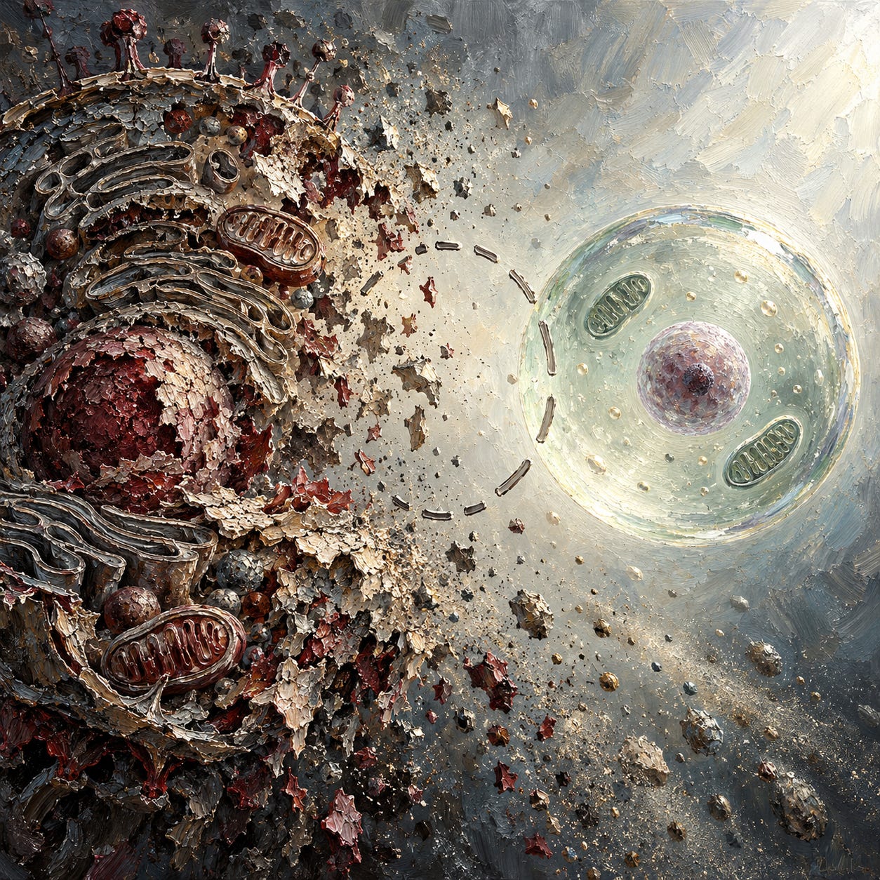
Author’s note: This essay engages with mainstream cell biology, virology, immunology, and genetics. Where that vocabulary appears, including viruses, immune system, antibodies, membrane receptors, ion channels, nuclear pores, and the central dogma, it operates in reporting voice, in quotation, in attribution, and in the official names of institutions and researchers. My own analytical voice operates in a different register, aligned with the terrain paradigm. Harold Hillman himself was not a terrain theorist. He was a mainstream physiologist whose objections operated inside the establishment’s own physics and solid geometry. His testimony is strongest when reported as his own, and the terrain reading emerges only after his case has been heard. Any errors of interpretation are mine.
The Picture in the Textbook
Open a biology textbook to the diagram of a cell. The cytoplasm is packed. Endoplasmic reticulum coils through the interior. A cytoskeletal lattice fills the remaining space. Golgi bodies stack and bud. Lysosomes and peroxisomes drift through the mesh. Ribosomes dot the reticulum. Molecular motors walk along microtubules carrying cargo. Receptors stud the membrane. Ion channels open and close in sequence. Mitochondria crowd the matrix, their inner membranes folded into cristae. Nuclear pores open and close in time with signaling events, carrying messenger RNA from nucleus to cytoplasm.
Now put a living cell under a light microscope.
What you see is a fluid of low viscosity. Nuclei and mitochondria move freely through it. Brownian motion is visible throughout. Streaming is visible. Nuclear rotation is visible. Material drifts from place to place by convection and diffusion. There is no mesh. There is no lattice. The cell is not solid.
Both pictures cannot be true at the same time of the same cell.
Harold Hillman spent fifty years asking which picture to believe.
Essay PDF Download (Free Subscribers)
Audio Deep Dive (Paid Subscribers Only)
Support This Work
The essays are free and will stay free. What paid subscribers get is the library that surrounds them: the full digital book catalogue, audio deep dives, archived, and embedded in each essay, and the Questions for Your Doctor, Before You Consent, and Package Insert series. Platinum Supporters also receive the digital edition of every paperback on the day it’s published.
Whichever tier suits, thank you for being here. If you’d like to become a paid subscriber, upgrade a tier, or gift a subscription to someone who might value the work, the options are below.
The Witness
Hillman held London University degrees in medicine and in physiology, and a doctorate in biochemistry.¹ He was Reader in Physiology at the University of Surrey from 1968 until he retired in 1995, and Director of the Unity Laboratory of Applied Neurobiology from 1970. He served as Medical Adviser to the Schizophrenia Association of Great Britain between 1990 and 1993. He published roughly one hundred and sixty full-length research papers and six books, on cytology, neurobiology, and resuscitation. He was the founding Editor-in-Chief of the journal Resuscitation from 1970 to 1985.
He was not a dissident by temperament. He was trained in the normal paradigms. He used electron microscopes. He ran subcellular fractionations. He conducted the experiments. What he discovered, and could not stop seeing, was that the methods his field relied on could not produce the information his field claimed to extract from them.
In 2011, long since stripped of institutional support, he published an open letter.¹ It was addressed to young research workers at the beginning of their careers in the early twenty-first century, before they had done work he considered methodologically compromised. The letter sets out, over roughly nineteen pages, why most of what cell biology has reported since the mid-twentieth century is methodologically indefensible.
The letter is not well known. The reason is instructive. We will come to it.
The First Objection: Thermodynamics
The second law of thermodynamics states that in a closed system any change in entropy must be accompanied by a change in free energy. Living systems are open. Energy and materials exchange freely between the organism and its environment. The rates and equilibria of chemical reactions inside a living cell are what they are because the cell is open, metabolizing, exchanging.
To study a cell, researchers extract it from the body, place it in a vessel, subject it to procedures, and measure what results. The vessel is not open. It is surrounded by media that conduct heat poorly: water, ice, plastic, glass, air, vacuum. Energy added to the system during preparation and measurement cannot dissipate quickly. The system heats, cools, shears, dilutes, denatures, recombines. The rates and equilibria of its reactions move away from those in the living animal.
Hillman cataloged the procedures routinely used in biochemical research and the effect each has on the entropy or free energy of the reactions being studied. The list in his paper runs to more than thirty entries. Killing the organism diffuses gradients. Tissue hypoxia reduces oxidative reactions. Post-mortem changes denature proteins. Cooling slows reaction rates. Slicing, homogenizing, macerating, and sonicating subject tissue to pressure, generate heat, and move enzymes, substrates, and activators from their original sites to other locations. Adding reagents dilutes the tissue and exchanges constituents. Washing extracts soluble material. Centrifugation generates friction and heat. Dialysis removes small molecules and ions. Precipitation changes solubility and reactivity. Drying concentrates all remaining constituents. Freezing and heating alter reaction rates and denature proteins. Chromatography and electrophoresis break bonds and heat tissue. Fixation inhibits enzymes and denatures proteins. Staining colors organelles and changes light absorption. X-rays and electron bombardment liberate heat and damage structures. Dissolution in powerful reagents denatures proteins and shifts pH.
Each of these steps changes what is being measured. In principle, none of them should be used to study living tissue unless and until the effects of each on the parameters of interest have been quantified. In practice, Hillman observed, no such control experiments have ever been published. Not for subcellular fractionation. Not for histology. Not for histochemistry. Not for electron microscopy. Not for binding studies.
In subcellular fractionation, the only popular control is to add up the enzyme activities in the final fractions at the end of a procedure and compare the total to the activity of the crude homogenate. If recovery is between sixty percent and one hundred and twenty percent, the experiment is considered satisfactory. If recovery is much lower, the activity in each final fraction is reported as a percentage of the total recovered rather than of the original homogenate, which conceals the loss. No experiment establishes that the properties of an organelle in a fraction reflect its properties in the living intact organism.
Hillman noted a further wrinkle on centrifugation. The g-force cited in a protocol refers to the center of the tube. The actual force varies with distance from the axis of rotation, so different parts of the tube are subjected to quite different quantities of energy. Different parts of the same homogenate therefore exhibit different chemical activities, which is unsurprising but is not what the experimenter is purporting to measure.
Across half a century, Hillman wrote, no biochemist told him that subcellular fractionation is governed by the second law; that control experiments have been published; that control experiments are not necessary; that the results of fractionation experiments do reflect the properties of intact tissue; or that the whole question is trivial. The silence was the evidence. Hillman reached for the Sherlock Holmes allusion: the dog had not barked.
The Second Objection: Solid Geometry
Electron microscopy delivers up to twenty-five times the resolution of light microscopy. When it was introduced to biology in the 1940s, it was greeted as a revelation. Structures previously invisible became visible. New names entered the vocabulary of the field. Textbooks were rewritten around what the new instrument appeared to show.
What the electron microscope actually requires is this. An animal is killed. The tissue is excised. It is fixed, or frozen. It is embedded. It is sectioned, often at thicknesses measured in nanometers. It is rehydrated. It is stained with heavy metal salts of osmium, tungsten, manganese, uranium, or lead. It is mounted. It is placed in vacuum. It is bombarded with electron beams.
What the microscope records is not the tissue. It is the deposit of heavy metal on the fixed, dehydrated section, in vacuum, under electron bombardment. If the heavy metal salts are not used for staining, virtually nothing is seen. Weakley’s 1972 handbook on electron microscopy carries an elegant illustration of this: an identical sample, with and without staining, in which the unstained version shows essentially nothing.²
The structures that then appear in electron micrographs include the trilaminar “unit membrane” first named by J.D. Robertson in 1959, in which cell membranes, nuclear membranes, and mitochondrial membranes all appear as a dark-light-dark sandwich of uniform thickness.³ The endoplasmic reticulum, an interior network. The Golgi body, previously described by Golgi in 1898. Networks of actin, tubulin, spectrin, and vimentin forming the cytoskeleton. Lysosomes and peroxisomes, each with their own portfolios of enzyme activities. Nuclear pores penetrating the nuclear membrane. Mitochondrial cristae, the shelves folded into the mitochondrial matrix. Thick and thin filaments composing the A and I bands of muscle, with small cross-bridges between them. Ribosomes, transmembrane molecules, membrane receptors, molecular motors, synapses, and synaptic vesicles.
Hillman identified most of these structures as artifacts of preparation. His argument is geometric before it is anything else.
Any genuine three-dimensional structure, when sectioned and photographed, should appear in a random distribution of orientations across a field of view. A long cylindrical structure cut transversely at one point in a field should appear longitudinally at another point and obliquely at a third. If every example of a particular structure appears in only one orientation, the structure cannot be three-dimensional. It must have appeared after the section was cut.
The unit membrane appears in electron micrographs almost always as a transverse section. Dark-light-dark lamellae of uniform thickness, cut normal to the plane of section, consistently across all membranes around cells, nuclei, mitochondria, and chloroplasts simultaneously. For this to be a real three-dimensional structure, every membrane in every organelle in every cell would have to be oriented normal to the plane of section at precisely the moment the microscopist chose to cut. Karl Deutsch pointed out the geometric impossibility in 1962.⁴ No one answered him.
The response from electron microscopists, when pressed, is that they publish the images in which membranes appear clearly, which happens to be the membranes cut transversely. Hillman accepted this as a candid answer and asked the next question. If selection explains the uniformity of orientation, how were the authors able to find transverse sections of all membranes in all organelles whenever they looked? For this to be true, the sections they cut must have contained membranes in random orientations, and other orientations must simply have been discarded as unclear. But in that case, the published literature should contain, somewhere, examples of membranes in random orientations in the same field at modest magnification. It does not.
The same geometric argument applies to the myelin lamellae, which appear only as two-dimensional sheets, and to the thylakoid membranes of chloroplasts, and to the mitochondrial cristae, and to the thick and thin filaments of muscle. In none of these cases do the published images present the structures in the variety of orientations that genuine three-dimensional structures must exhibit.
The cytoplasm argument closes the circle. If all the structures identified by electron microscopy actually existed in a living cell at the quantities implied by electron micrographs, the cell would be nearly solid. The cytoskeleton alone, if present as drawn, would prevent movement. The endoplasmic reticulum would obstruct diffusion. The sarcoplasmic reticulum would obstruct intracellular motion. Yet living cells observed under light microscopy show a fluid of low viscosity in which nuclei and mitochondria move freely and in which Brownian motion, streaming, convection, nuclear rotation, phagocytosis, pinocytosis, vacuolation, mitosis, and muscle contraction all occur. The two pictures are not compatible. If the light-microscopy picture is accurate, most of the electron-microscopy picture cannot be.
The techniques have been refined in the decades since. Cryo-electron microscopy freezes samples in vitreous ice to try to minimize some fixation and dehydration artifacts. Super-resolution light microscopy pushes beyond the optical diffraction limit by other physical means. Atomic force microscopy maps surfaces with a sharp probe. Each still requires material taken from the living organism and prepared on grids, in vacuum, under beams or probes that bring energy to a system that cannot remain open to its environment. The thermodynamic objection applies to all of them. The geometric constraint applies to whatever any of them shows. The methods have improved. The problem Hillman named remains in place.
Hillman accepted two structures as present in the cytoplasm of living cells. Nuclei, containing nucleoli. Mitochondria. Nothing else.
The Catalog
Hillman’s 2011 paper lists twenty-seven structures that he identified as artifacts of preparation, with the likely origin of each. Several deserve naming here, both because of their status as textbook orthodoxy and because of what their artifactual status means for everything built on them.
The unit membrane is a two-dimensional artifact produced after sections were cut.
Ion channels: only one, the sodium acetylcholine channel, has been modeled, by Kistler and colleagues in 1982.⁵ The others are inferences from electrophysiological measurements that could be explained in other ways.
The endoplasmic reticulum and the cytoskeleton are precipitates of staining reagents in dehydrated cytoplasm. They are not seen in three dimensions at equal frequency and would, if present, prevent the intracellular movements observed in living cells.
Lysosomes were conceived after a subcellular fraction enriched with hydrolytic enzymes was separated. The structure was then “seen” in electron micrographs to match the biochemistry. Peroxisomes originated the same way, from a fraction enriched in roughly thirty enzymes including peroxidases and catalases.
Membrane receptors and transmembrane molecules arise from the pharmacological assumption that drugs act only by binding to specific proteins, and from the biochemical assumption that ions and amino acids require macromolecules to cross membranes. Neither has been observed in living cells by any form of microscopy.
Synapses and synaptic vesicles are precipitates of silver and heavy metal salts near nerve cell bodies, giving the appearance of specialized junctions. Synaptic vesicles appear in electron micrographs with suspiciously uniform diameters and without visible stalks.
Mitochondrial cristae are the dehydrated matrix of the mitochondrion appearing as folded shelves. The outer and inner mitochondrial membranes are, Hillman argued, a single membrane appearing as two lines because of the limits of resolution.
Nuclear pores are cracks in the nuclear membrane produced during preparation. The hypothesized passage of messenger RNA across them, central to the molecular biology of gene expression, has never been observed. It is a hypothesis built around a structure that may itself be a crack.
Thick and thin muscle filaments and the cross-bridges between them do not conform to three-dimensional geometry when examined across published electron micrographs. Muscle in life is a gel. When dehydrated for electron microscopy, it precipitates as fibers.
The catalog is longer. The pattern is uniform. Structures claimed to be the architecture of the cell, taught as such in every textbook, cannot be reconciled with what living cells actually show when examined by techniques that leave them alive.
The Dog That Did Not Bark
In 1964, before a meeting of the British Physiological Society at Mill Hill, Hillman was warned by the chairman, William Feldberg, that his paper would be attacked. He offered to withdraw temporarily if his opponent would undertake to repeat his experiments. The opponent refused. The paper was presented. Hillman was attacked by the pharmacologist Gustav Born and asked five questions, three of which he had already answered in his presentation. Of roughly two hundred physiologists present, about fifteen voted against publication of the abstract, four voted for, and the rest abstained.
The following Monday, the biophysicist J.A.V. Butler telephoned him to say that, in his view, the rejection had ruined Hillman’s chances of ever being appointed to a permanent academic post in Britain.
Butler was substantially correct.
From 1972 onwards, after the publication of his first book cataloging methodological failures in biochemical techniques, Hillman received no further public funding for his research. His salary as Reader in Physiology at Surrey continued, until the university attempted to close his Unity Laboratory. The grounds given were that his research was expensive, his scientific views were unpopular, and he had been unable to attract external funds. He produced documents showing his laboratory was the cheapest in the faculty and that seventy percent of academic staff had similarly failed to attract outside funding. A question was asked in Parliament about whether the cut would limit the expression of unpopular views. The Association of University Teachers backed him. The Council and Senate voted to close the laboratory and remove his tenure. He kept working full-time until retirement at sixty-five, supported by a children’s charity and by the pharmaceutical executive David Horrobin.
He applied for a chair after twenty-two years as Reader. The application was turned down. The Vice-Chancellor told him the reason was that a Fellow of the Royal Society had informed him that Hillman’s views were considered controversial among biologists.
He wrote to the pioneers of the views he contested. Christian de Duve, who proposed lysosomes. Hugh Huxley, who proposed the sliding filament model. Bernard Katz, who proposed the quantal release of neurotransmitters at synapses. Keith Porter, the pioneer of electron microscopy in cell biology. None would meet him. Hans Krebs met him. Ernest Chain met him. J. David Robertson, who named the unit membrane, met him. Andrew Huxley met him. Most senior figures declined on various grounds. One wrote back saying that more heat than light would be generated if they met.
He was prevented from presenting papers at the International Society for Neurochemistry, the European Society for Neurochemistry, the German Physiological Society, and the British Physiological Society, where he had been a member for over forty years. No reasons were given. Sometimes he was offered a poster, which he refused as inadequate for the material.
In 2010, The Biologist magazine published a short article of his doubting the validity of subcellular fractionation and electron microscopy.⁶ The editor was rebuked by the chairman of the Society of Biology and, Hillman believed, replaced. The Society of Biology and the Royal Microscopical Society jointly authorized Professor Christopher Hawes of Oxford Brookes University to write a response.⁷ Hillman considered it extraordinary that two learned societies should give their official support to one side in a scientific disagreement. He did not feel Hawes had addressed the points he had made. The Biologist refused him the right of reply.
At every lecture he gave, in Britain, Europe, North America, Israel, Thailand, and Australia, members of the audience approached him privately to say they agreed with him. He asked each of them if they were prepared to express the same views in public. The replies named stages of a career. One was typical: “No, no, I am afraid that I cannot do so as I’m completing my Ph.D.” Others cited lectureship applications, grant proposals in review, and chair applications in progress. Two electron microscopy technicians who prepared illustrations for one of his books declined to be acknowledged by name. “It would be more than our careers would be worth.”¹
Across half a century, no biochemist published control experiments addressing the thermodynamic objection. No electron microscopist addressed the geometric objection on its own terms. No textbook author modified the diagrams. No pioneer agreed to repeat the experiments. The dog did not bark. The structures remained in the textbooks because they had been in the textbooks, and the students who read the textbooks became the professors who wrote the next edition. What rests on them collapses with them.
Cascade: Virology
A virus particle, as presented to the public and in the literature, is an electron microscope image. The particle has a shape. It has a diameter in nanometers. It has spikes, or a lipid envelope, or a crystalline capsid structure. The images are reproduced in textbooks, press releases, and public health announcements.
The particle is produced as follows. A clinical sample from a sick person, which may be a nasopharyngeal swab eluate, a lung lavage, or a tissue biopsy, is inoculated into a cell culture. The culture medium is adjusted to deprive the cells of certain nutrients, and antibiotics are added. The cells degrade. Material is extracted from the degraded culture, centrifuged, fixed, embedded, sectioned, stained with heavy metal salts, placed in vacuum, bombarded with electrons, and photographed.
Each of these procedures is one Hillman identified as changing the properties of the sample grossly and significantly, without published controls to quantify the change. The cell culture conditions themselves degrade cells. Starving and poisoning a cell culture produces cellular breakdown products. Centrifugation generates heat and selects fractions based on density. Fixation and dehydration denature proteins. Heavy metal staining produces deposits whose relationship to any underlying biological structure is governed by the chemistry of the stain, not by the structure of the sample. Electron bombardment liberates further heat and x-rays.
The virological establishment has not published control experiments in which the same procedures are performed on uninoculated cultures to establish whether the particles seen in inoculated cultures could have been produced by the procedures alone. One notable exception sits in the paper that founded modern cell-culture virology. In 1954, John Enders and Thomas Peebles reported their isolation of what became known as the measles virus. In the same paper they recorded that an uninoculated monkey kidney cell culture produced cytopathic changes they “could not distinguish with confidence from the viruses isolated from measles.”⁸ The admission was in the founding paper. It was not followed up.
Stefan Lanka subsequently took the measles claim to court after offering a cash prize for a single scientific publication demonstrating the virus’s existence. The case ran from 2012 to 2017 and reached Germany’s Federal Court of Justice, the Bundesgerichtshof, which ruled in his favor on the question of whether the papers submitted met the burden of proof demanded.⁹ During the trials Lanka commissioned independent German laboratories to perform the control experiments the field had not done. The results replicated what Enders had conceded sixty years earlier. Cytopathic effects appeared in cultures that had not been inoculated with any patient sample, driven by the starvation and antibiotic poisoning imposed on the cells by the culture conditions themselves. Mark Bailey and Sam Bailey have since documented the same methodological failure across the full history of virology.¹⁰
Hillman’s objection to electron microscopy generally cuts beneath the virological case specifically. If the method cannot distinguish real cellular structures from preparation artifacts, it cannot distinguish what are claimed to be virus particles from preparation artifacts either. The genomic sequences assembled from short reads amplified by PCR cannot repair this. A sequence derived from material that was never purified in whole-organism form is a sequence derived from a mixture. The assembly is a computational construct.
The “virus” was never seen by any method that could show it was there.
Cascade: The Spike Protein
The spike protein, as presented in the vaccine era, is a specific molecule with a specific structure, a specific binding behavior, and a specific set of claimed physiological effects. It appears in diagrams as a crown of protrusions on the surface of a sphere.
The protein was never isolated from a sick person. Its structure was modeled on a computer from genetic sequences assembled by PCR-based methods out of material that was never purified in whole-organism form. The protein used in binding studies, in hemagglutination assays, and in toxicity experiments is a laboratory product manufactured from the hypothesized sequence.
Hillman’s work bears on this chain at two points. First, the sequences themselves derive from material viewed through electron microscopes and processed through techniques he identified as unable to distinguish real cellular structures from artifacts of preparation. The “virus” from which the spike sequence was derived is itself a construct of unverified methods. Second, the receptors to which the modeled spike is said to bind, including ACE2, are membrane proteins of a class Hillman identified as never observed in living cells by any form of microscopy. The entire picture of a spike protein binding a membrane receptor to initiate cellular entry is a cartoon built from structures neither end of which has been observed in intact tissue.
What the injection products deliver is a different question. The lipid nanoparticles are real substances in the vial, measurable by chemistry. So are the modified mRNA, the aluminum where present as adjuvant, the polyethylene glycol, and the DNA fragments detectable in the final product. These are physical ingredients. Their effects on tissue can be studied.
What the transfected cells then produce is less certain than the marketing suggests. The output of the injection has been named “spike protein” because that was the sequence encoded. Whether what the cells actually express corresponds to the modeled structure, binds to anything modeled as a receptor, or causes the effects attributed to it in the discourse are questions that rest on the same methodological foundations Hillman spent fifty years dismantling.
The spike protein as a specific entity with specific causal properties is a construct downstream of a construct downstream of a method Hillman showed cannot do what it claims.
Cascade: Immune System Machinery
The conventional account of immunity requires a specific architecture. The cell membrane is studded with receptors that recognize specific molecular patterns. Antigens bind to those receptors with high specificity. Signaling cascades propagate through membrane-bound and cytoplasmic proteins. B cells produce antibodies, Y-shaped proteins secreted into the circulation, which bind foreign molecules and mark them for destruction. T cells recognize antigens presented on major histocompatibility complexes. Macrophages engulf and digest tagged material. Natural killer cells identify and lyse infected cells by means not fully specified.
The architecture requires receptors on the membrane. Hillman identified membrane receptors as structures never seen in life by any form of microscopy. The pharmacological assumption that drugs act only by binding to receptors generated the concept; the concept was then “seen” in subcellular fractions enriched by binding assays. The architecture requires transmembrane molecules shuttling signals across the lipid bilayer. Hillman identified these as never observed. The architecture requires ion channels gating the passage of charged species. Only one has been modeled.⁵
What medicine calls natural antibodies have never been purified and characterized as discrete molecules in human serum. The antibodies used in research and diagnostics are a different kind of object. In 1975, Georges Köhler and César Milstein developed a technique for fusing a mouse’s B cell with a myeloma, a cancerous plasma cell that multiplies indefinitely. The hybrid cell, called a hybridoma, produces a single antibody variant at scale. The 1984 Nobel Prize in Physiology or Medicine was awarded for this discovery.¹¹ The products are the monoclonal antibodies that populate the research literature. They are laboratory-manufactured chimeras of mouse and cancer, produced to a specification.
Even by the measures of the field that produced them, their specificity is in question. Clifford Saper, Harvard neurologist and Editor-in-Chief of the Journal of Comparative Neurology from 1994 to 2011, kept receiving distressed submissions from authors whose antibodies had stained tissue in animals that lacked the target entirely. In 2003 he and Paul Sawchenko required authors to document the methods used to validate specificity, and gave the unvalidated reagents a name: “magic antibodies.”¹² A dozen years later, Monya Baker’s 2015 analysis in Nature reported that something close to half of commercial antibodies on the market fail basic validation tests, and traced a substantial share of biomedical research’s reproducibility crisis directly to this failure.¹³
What is measured in a routine antibody test is the binding of proteins in serum to a prepared substrate under laboratory conditions, inferred from a color change, a luminescence, or a radioactive count. The interpretation of the measurement as the presence of a specific immunoglobulin targeting a specific antigen rests on specificity assumptions that remain difficult to establish by any method capable of observing the binding directly.
The architecture requires the “immune system” as an integrated functional whole, with its memory, its surveillance, its self-nonself recognition. You cannot open a body and point to it. What exists anatomically is the lymphatic system, the fascial network, and the bone marrow. These are organs of circulation, filtration, and cell production. The “immune system” is a conceptual overlay. It was built to explain why germ theory’s predictions fail at the individual level. If a specific germ caused a specific disease, exposure should produce the disease uniformly. It does not. The “immune status” of the host was invoked to patch the discrepancy.
If the membrane receptors, transmembrane molecules, and ion channels through which the immune machinery is supposed to operate are not present as described, and if the natural antibodies on which the humoral arm depends have never been isolated as discrete molecules, and if the manufactured antibodies on which modern research runs are chimeras of mouse and cancer whose specificity the field itself cannot reliably establish, then what medicine calls immunity requires a different account. The account that remains is the one the terrain tradition has given for a century and a half. Cells of the lymphatic network perform waste management on cellular debris and other foreign material. The body does its own cleansing and repair, not warfare on invaders. The terrain decides what manifests.
Cascade: Genetics and the Central Dogma
The central dogma of molecular biology, in the form Francis Crick articulated in 1958 and amended in 1970, states that genetic information flows from DNA to RNA to protein. The mechanism requires that messenger RNA, transcribed from DNA in the nucleus, cross from the nucleus to the cytoplasm, where it is translated by ribosomes into protein. The crossing happens, in the standard account, through nuclear pores: specialized gated channels penetrating the nuclear membrane.
Hillman identified nuclear pores as cracks in the nuclear membrane produced during preparation for electron microscopy. The passage of messenger RNA across them has never been observed in living cells. It is a hypothesis built around a structure that may itself be an artifact of the fixation and sectioning that generated the image in which the structure appears.
Ribosomes, in Hillman’s account, were conceived after a subcellular fraction containing RNA was separated. The particles seen by electron microscopy in sections or in fractions were then named ribosomes to match the biochemistry. Whether discrete particles of the hypothesized structure exist in intact living cells, performing the translation function attributed to them, is a question that has not been answered by any method that observes living cells.
The DNA molecule as described by Watson and Crick exists. Nucleic acids are real chemical substances with known structures, extractable from tissue and characterizable by crystallography. What the terrain reading contests is the central dogma’s physical mechanism, which requires transport through structures Hillman identified as artifacts, through particles Hillman identified as subcellular constructs, into a cytoplasm Hillman identified as largely free of the organelles claimed to populate it.
The Human Genome Project set out to identify the genes that encode human proteins. Mainstream estimates before completion ranged above one hundred thousand. The project found approximately twenty thousand.¹⁴ The expected correspondence between genes and complex biological traits has not materialized. Genome-wide association studies have failed to produce the explanations for complex traits they were designed to deliver. The “gene for X” model has failed repeatedly for complex traits and chronic diseases.
What remains of the central dogma as a complete causal theory of biology is less than the public presentation suggests. What remains in clinical practice is the “genetic predisposition” category: a diagnostic placeholder used to foreclose investigation of toxicological, nutritional, electromagnetic, and psychological causes, and to justify lifelong pharmaceutical management of symptoms whose actual origins lie elsewhere.
The physical mechanism by which DNA is said to cause disease, mediated through structures Hillman identified as artifacts, has not been demonstrated. The clinical application that depends on the mechanism is accepted anyway, because the alternative is to look at what is actually making people sick.
The Missing Diseases
Hillman closed his 2011 paper with an observation he presented as a hypothesis and that reads now as a verdict.
Modern medical research, he suggested, could be divided into three kinds. The first is serendipitous. Penicillin, lithium for bipolar disorders, neuroleptics for schizophrenia, steroids for skin conditions. Important findings arrived by accident. The second is applied technology. Intensive care units, cardiac surgery, keyhole surgery, tissue transplantation. Advances flowed from engineering rather than from basic science. The third kind is basic research into the genesis of disease: cancer, multiple sclerosis, schizophrenia, Alzheimer’s dementia, motor neuron disease, muscular dystrophy. Research intended to identify the mechanisms whose disarray initiates these conditions, so that rational interventions might be designed.
“It is absolutely remarkable,” Hillman wrote, “how unsuccessful this sort of research has been.”¹
He put forward the hypothesis that the poor quality of cell biology in the twentieth century was the reason. If the methods used to investigate living tissue cannot produce accurate information about living tissue, the field cannot find the chemical changes that initiate disease. The cost of the failure, Hillman noted, had been high.
Place this verdict next to what the terrain tradition has argued since Antoine Béchamp and Claude Bernard. Disease arises from the internal environment of the body. The four insults are toxic exposure, nutritional deficiency, electromagnetic disturbance, and psychological strain. These insults operate in living bodies, in whole organisms, through fluids and tissues and metabolic cycles that are open to the environment and that cannot be captured in a vessel without changing their properties.
A cell biology built on dead, dehydrated, metal-stained tissue cannot find the causes that operate in living bodies. The biochemistry built on homogenization and centrifugation cannot find them either. Pharmacology built on membrane receptors never observed in life cannot address them. Molecular biology whose central mechanism runs through what Hillman identified as cracks in the nuclear membrane cannot explain them. Virology, whose observational foundation is electron microscopy of degraded cell cultures, cannot distinguish them from the artifacts the field has been studying all along.
Eighty years of failed basic research on the origins of chronic disease traces to a methodological structure incapable of producing the answer. Hillman identified the structure through the physics and the geometry. The terrain tradition had already identified what actually makes people sick, from the clinical and the historical side.
The cell biology taught in textbooks is a picture of what dead tissue looks like after particular preparation for a particular form of microscopy. The living cell has turned out to be simpler and more fluid than the textbook picture, and far more of what matters in it remains unseen than the field admits. Everything built on the textbook picture, including the virological account of infection, the immunological account of defense, the genetic account of susceptibility, and the pharmacological account of therapy, rests on something that was never there. The sick are managed with interventions designed against this picture, which is one reason the sick keep getting sicker.
Hillman addressed his open letter to the young, who had not yet published compromised work and who might choose different methods. He died on 5 August 2016, eleven days short of his eighty-sixth birthday. The open letter was then five years old and had changed nothing. In 2026, ten years after his death, the textbook pictures remain unchanged. The research funding still flows to projects built on them. The structures Hillman identified as artifacts continue to populate every introductory biology course.
What makes people sick operates in whole, living bodies: in the toxins they absorb, the food they do not get, the fields they stand in, the lives they must endure. These are the questions the methodology Hillman described cannot ask. The terrain tradition could ask them because it never left the whole living body in the first place.
Everything else has been looking at dust.
How to Explain It to a Six-Year-Old
Imagine you have a snowman in the garden. You want to know how snowmen work on the inside, so you decide to study it. But to study it properly, you take it inside and put it on the kitchen table. The warm kitchen melts it. The water runs everywhere. You scoop up some of the puddle and dry it out, and now you have some flakes of dust. You put the dust under a magnifying glass. You draw a picture of what you see. You call that picture a snowman.
But the picture of the dust is not what a snowman is. A snowman is cold and round and standing in the garden. The picture of the dust is what happens to a snowman after you take it inside, melt it, and dry it out. If you want to know what makes a snowman stand up or fall over, looking at the dust will not tell you. You have to go back outside and look at the snowman.
Scientists made the same mistake for a long time with the tiny parts of our bodies. They took the parts out of the body, where they were alive and moving. They dried them. They put metal on them. They made pictures of what was left. Then they said the pictures showed how the body works.
The pictures showed what happens to tiny body parts after you take them out, dry them, and put metal on them. The pictures did not show the body.
That is why when people get sick, the doctors often cannot say why. They have been looking at the dust.
Additional Sources
Bailey, M. and Bailey, S. Various essays, interviews, and books on virology methodology, including A Farewell to Virology and The Final Pandemic: An Antidote to Medical Tyranny. Available at drsambailey.com.
Cowan, T. (2020). The Contagion Myth: Why Viruses (Including “Coronavirus”) Are Not the Cause of Disease. Skyhorse Publishing.
Engelbrecht, T. and Köhnlein, C. (2021). Virus Mania: Corona/COVID-19, Measles, Swine Flu, Cervical Cancer, Avian Flu, SARS, BSE, Hepatitis C, AIDS, Polio, Spanish Flu. How the Medical Industry Continually Invents Epidemics, Making Billion-Dollar Profits at Our Expense. 3rd edition.
Gober, M. with Bailey, S., Bailey, M., Lanka, S., et al. (2023). An End to Upside Down Medicine: Contagion, Viruses, and Vaccines, and Why Consciousness Is Needed for a New Paradigm of Health. Waterside Productions.
Lester, D. and Parker, D. What Really Makes You Ill? Why Everything You Thought You Knew About Disease Is Wrong.
Martin, B. (ed.) (1996). Confronting the Experts. State University of New York Press. Includes Hillman’s “What price intellectual honesty?” essay with full bibliography of his work to 1996.
Roytas, D. (2024). Can You Catch a Cold? Untold History and Human Experimentation.
Shelton, H.M. Natural Hygiene: Man’s Pristine Way of Life; collected articles and books.
Tilden, J.H. Toxemia Explained: The True Interpretation of the Cause of Disease.
Williams, U. and Bailey, M. (2022). Terrain Therapy.
In Print
The Unbekoming library is available in paperback, printed to order through Lulu and shipped worldwide. The shelf begins with the paradigm question underneath everything else — No Virus, the isolation problem, the collapse of virology’s foundational claims, and a disease-by-disease reappraisal — and moves through the suppressed compounds mainstream medicine set aside: The DMSO Book, Chlorine Dioxide: The Forbidden Remedy, The Iodine Book, and The Hydrogen Peroxide Book. Two more recover what’s still on the kitchen shelf: Baking Soda and The Castor Oil Book. Two more recover the minerals modern soil, water, and processing quietly stripped from the diet: The Magnesium Handbook and The Boron Book. Sitting alongside these is No Contagion, co-authored with Jamie Andrews — the case against germ theory itself, catalogued through 258 failed contagion experiments.
The critique books cover what medicine, dentistry, psychiatry, and veterinary practice have become. The Unvaccinated treats the completely unvaccinated as a comparison group across twenty chapters and five appendices. Medicalized Motherhood follows a woman through 123 documented interventions from teenage pill to postpartum discharge. Drilling for Profit treats cavities, gum disease, and crooked teeth as the dietary problem they are. What Your Vet Can’t Tell You applies the same critique to pets. Escape from Psychiatry documents the fabrication of the DSM and the specific damage of every major psychiatric drug class. The Vitamin K Injection covers what happens in the first hours of a newborn’s life.
The full shelf is at lulu.com/spotlight/unbekoming. A physical book reaches the person a Substack post never will — the skeptical relative, the friend who won’t click a link but might open a book, the visitor whose eye lands on a coffee table. Buy one to keep, and one to give away.
References
Hillman, H. (2011). Cell Biology is Currently in Dire Straits. Unity Laboratory of Applied Neurobiology, 76 Epsom Road, Guildford, Surrey, GU1 2BX, U.K. Latest version dated 29 June 2011.
Weakley, B. (1972). Beginners Handbook in Biological Electron Microscopy. Churchill-Livingstone, London.
Robertson, J.D. (1959). The ultrastructure of cell membranes and their derivatives. In The Structure and Function of Subcellular Components. Cambridge University Press, Cambridge.
Deutsch, A. (1962). Artifacts, the great problem of electron microscopy. Wissenschaftliche Zeitschrift der Ernst Moritz Arndt Universität Greifswald, XI, Math. Natur. Reihe, 2.
Kistler, J. et al. (1982). Structure and function of an acetylcholine receptor. Biophysical Journal, 37, 371-383.
Hillman, H. (2010). Two problems with cell biology and what should be done about them. The Biologist, 57, 42-46.
Hawes, C. (2010). No problems in cell biology. The Biologist, 57.
Enders, J.F. and Peebles, T.C. (1954). Propagation in tissue cultures of cytopathogenic agents from patients with measles. Proceedings of the Society for Experimental Biology and Medicine, 86(2), 277-286.
Bundesgerichtshof (Federal Court of Justice of Germany), Case VI ZR 36/16, judgment of 16 February 2017 (Lanka v. Bardens, final appeal). Reversing the Oberlandesgericht Stuttgart decision of 16 February 2016. Trial and related control-experiment documentation reviewed in Lanka’s published writings and in works by the Baileys.
Bailey, M. (2023). The Final Pandemic: An Antidote to Medical Tyranny; and Bailey, S. and Bailey, M., A Farewell to Virology and collected works at drsambailey.com.
Köhler, G. and Milstein, C. (1975). Continuous cultures of fused cells secreting antibody of predefined specificity. Nature, 256(5517), 495-497. Nobel Prize in Physiology or Medicine awarded 1984 to Köhler, Milstein, and Niels K. Jerne.
Saper, C.B. and Sawchenko, P.E. (2003). Magic peptides, magic antibodies: Guidelines for appropriate controls for immunohistochemistry. Journal of Comparative Neurology, 465(2), 161-163. See also Saper, C.B. (2005). An open letter to our readers on the use of antibodies. Journal of Comparative Neurology, 493(4), 477-478.
Baker, M. (2015). Reproducibility crisis: Blame it on the antibodies. Nature, 521, 274-276.
International Human Genome Sequencing Consortium (2004). Finishing the euchromatic sequence of the human genome. Nature, 431, 931-945.
Congratulations on this excellent article, which invites reflection on a fundamental question: to what extent might the scientific methods used to study living cells shape—or even distort—our understanding of disease?
I find Hillman’s perspective on the limitations of studying tissues outside their biological context particularly compelling. An organism is not merely a collection of isolated cells; it is a dynamic system in which metabolism, nutrition, the environment, and adaptive mechanisms constantly interact.
Exposing the fallacy of the classic cellular model—that static, neatly packaged "textbook" construct which never existed within the dynamic reality of living tissue—effectively dismantles the very foundations upon which modern pathology has been built.
Harold Hillman’s critique, rooted in the physics and thermodynamics of intact tissue, aligns brilliantly with broader critiques of mechanistic reductionism. When living matter is isolated, stained, fixed, and subjected to ultracentrifugation, we are not uncovering the true nature of disease; rather, we are creating a laboratory artifact. Pathology does not arise from microscopic glitches within "ghost cells" analyzed in isolation; it emerges from a systemic disruption of the entire organism.
This conclusion resonates with a tradition—championed by figures such as Antoine Béchamp and Claude Bernard—which posits that disease arises from the body’s internal environment. Four primary triggers are involved: exposure to toxins, nutritional deficiencies, electromagnetic disturbances, and psychological stress. These factors exert their influence on living, whole organisms—through fluids, tissues, and metabolic cycles that are inherently exposed to the environment and cannot be confined within a laboratory vessel without fundamentally altering their properties.
The root causes of human illness operate within living, intact bodies: in the toxins absorbed, the essential nutrients missed, the environments inhabited, and the life circumstances one is forced to endure. These are the questions that the methodology described by Hillman cannot pose. The "terrain" tradition, however, could pose them because, from the very beginning, it never strayed from the living body in its entirety.
Returning to look at the living body—at the "terrain," the fluidity of the internal environment, and the actual conditions of existence (nutrition, toxins, stress, and surroundings)—is the only way to rescue physiology from its methodological dead end.
****THAT IS THE FUNDAMENTAL DIFFERENCE BETWEEN PHARMACOLOGICAL REDUCTIONISM AND THE BIOLOGY OF THE "TERRAIN." THANK YOU FOR ARTICULATING THIS PROFOUND CRITICAL REVIEW.****
Against the backdrop of Hillman’s framework—and a tradition of thought tracing back to Antoine Béchamp and Claude Bernard—we arrive at the subject of vaccines. Vaccines operate within a mechanistic paradigm: they attempt to isolate a response against a specific antigen in a laboratory setting, based on the assumption that the body functions as a collection of independent parts that can be "trained" through artificial external stimuli (such as toxins, adjuvants, and viral fragments in a sterile medium).
In reality, the strength of the immune system is not a switch that can be flipped by an injection; rather, it is a direct reflection of the health of the entire organism. It depends on a clean internal environment, optimal cellular nutrition, the integrity of tissues, the absence of accumulated toxins, and the body’s ability to maintain its dynamic equilibrium (homeostasis) in relation to its surroundings.
A biological "terrain" that is strong, well-nourished, and respected in its entirety does not need to be "patched" from the outside through the imposition of synthetic antigens; instead, it responds to life’s challenges in a natural, systemic, and coherent way. To conflate the artificial induction of antibodies in the blood serum with genuine immunological robustness is to overlook how life truly functions within a living, integrated organism.
Harold Hillman’s analysis of the methodological limitations of cell biology—and the reductionist approach typical of laboratory research—helps us understand the structural problems inherent in vaccines, viewed from a strictly physiological and thermodynamic perspective:
• The dogma of cellular and tissue isolation: Vaccines are designed and evaluated under the assumption that cells and immune components act as independent, static units that can be "stimulated" in a linear fashion within a laboratory vessel. However, as Hillman demonstrated through his study of living tissue, bodily fluids and cells actually operate within a continuous, low-viscosity, and highly dynamic environment. Introducing foreign agents, adjuvants, or synthetic nucleic acids directly into tissues alters biophysical properties that traditional laboratory models can never adequately replicate or anticipate.
• The fallacy of equating antibodies with immunity: The official paradigm measures the success of a vaccine based on a single, isolated marker—the production of serum antibodies as detected in a blood test—while ignoring the complexity of the innate and systemic immune system. A high antibody titer does not equate to a robust biological terrain or the health of the organism as a whole, just as a fixed histological specimen is not the same as living tissue.
• Assault on the internal environment (the "terrain"): From the perspective of "terrain-based" medicine, any intervention that forces a systemic inflammatory response—through the direct inoculation of toxins, metallic adjuvants (such as aluminum salts), or genetic platforms—bypasses the body’s natural defensive barriers (mucous membranes, skin, microbiota, and the liver and kidney detoxification systems). By disregarding the broader environment and the actual underlying triggers—such as nutrition, accumulated toxic burden, and metabolic stress—an artificial load is imposed upon deep tissues, disrupting homeostasis rather than strengthening it.
--